. 1999 Feb;19(2):1410-5.
doi: 10.1128/MCB.19.2.1410.

Essential role of the dynamin pleckstrin homology domain in receptor-mediated endocytosis

Affiliations

Essential role of the dynamin pleckstrin homology domain in receptor-mediated endocytosis

M Achiriloaie et al. Mol Cell Biol. 1999 Feb.

Abstract

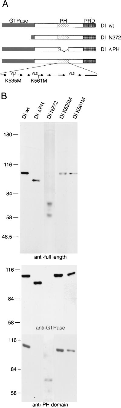
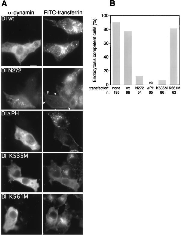
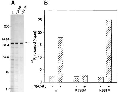
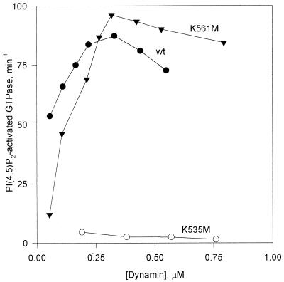
Pleckstrin homology (PH) domains are found in numerous membrane-associated proteins and have been implicated in the mediation of protein-protein and protein-phospholipid interactions. Dynamin, a GTPase required for clathrin-dependent endocytosis, contains a PH domain which binds to phosphoinositides and participates in the interaction between dynamin and the betagamma subunits of heterotrimeric G proteins. The PH domain is essential for expression of phosphoinositide-stimulated GTPase activity of dynamin in vitro, but its involvement in the endocytic process is unknown. We expressed a series of dynamin PH domain mutants in cultured cells and determined their effect on transferrin uptake by those cells. Endocytosis is blocked in cells expressing a PH domain deletion mutant and a point mutant that fails to interact with phosphatidylinositol 4,5-bisphosphate [PI(4,5)P2]. In contrast, expression of a point mutant with unimpaired PI(4,5)P2 interaction has no effect on transferrin uptake. These results demonstrate the significance of the PH domain for dynamin function and suggest that its role may be to mediate interactions between dynamin and phosphoinositides.


Figures

FIG. 1
FIG. 1
Characterization of expressed dynamin mutants. (A) Diagram of the expressed mutant dynamin constructs. The PH domain structural elements (8) are shown in the bottom expansion, in which the arrows represent β-sheet strands, and the rectangle represents an α-helix. VL, variable loop. (B) Immunoblots of transfected Cos cell extracts, obtained with antidynamin antibodies raised against intact rat brain DI (anti-full length), against a synthetic peptide corresponding to residues 607 to 624 (anti-PH domain), and against residues 45 to 358 (anti-GTPase). The construct transfected is indicated above each lane. Numbers on the left are molecular masses, in kilodaltons. The 60-kDa band recognized by the antibodies in DI N272 preparations is probably a degradation product. wt, wild type.
FIG. 2
FIG. 2
Effects of DI mutants on transferrin internalization. (A) Immunofluorescence images of cells. Left panels show the transfected cells identified by staining with antiserum against full-length DI (α-dynamin). Right panels show the fluorescence signals from cells which have internalized FITC-transferrin; arrowheads point to the cell transfected with DI N272. Bar, 10 μm. (B) Quantitative assessment of the endocytic poisoning potential of each construct. Cells considered endocytosis incompetent lacked any FITC-transferrin spots. wt, wild type.
FIG. 3
FIG. 3
PI(4,5)P2 stimulation of the GTPase activity of dynamins expressed in Sf9 cells. (A) Coomassie blue-stained gel of Sf9 cell extracts used for GTPase assays. The arrow indicates dynamin. wt, wild type. Numbers on the left are molecular masses, in kilodaltons. (B) GTPase activities of Sf9 cell extracts containing wild-type (wt) or mutant dynamins measured in the absence or presence of 5 μM PI(4,5)P2. Assays were carried out in buffer A containing additionally 0.1 M NaCl for 5 min. Dynamin concentrations (0.44 μM wt DI, 0.57 μM DI K535M, and 0.53 μM K561M) were estimated by densitometric scanning of Coomassie blue-stained gels (as in panel A), using electrophoresed bovine serum albumin to generate a standard curve. Typical GTPase activities of mock-infected Sf9 cell extracts were one-third to one-half of the basal values of dynamin-expressing extracts.
FIG. 4
FIG. 4
Specific PI(4,5)P2-stimulated GTPase activities of wild-type (wt) and mutant dynamins as a function of dynamin concentration. Specific activities were calculated by dividing moles of Pi released per minute by the dynamin concentration in each assay. Activities in the absence of PI(4,5)P2 were subtracted from each data point. Experiments were performed in buffer A containing 0.1 M NaCl. The PI(4,5)P2 concentration was 5 μM.
FIG. 5
FIG. 5
Interactions of dynamin mutants with GST-Grb2 and microtubules. (A) GTPase activation by GST-Grb2. The assays were performed in buffer A containing 0.1 M NaCl. (B) Effect of microtubules (MT) on GTPase activity. Assays were carried out in buffer A without added salt. (C) Binding of expressed dynamins to microtubules. Wild-type (wt) and mutant dynamins were subjected to a cosedimentation assay with microtubules (5 μM tubulin dimer). Means and standard errors of triplicate measurements are shown. Concentrations of wt DI, DI K561M, and DI K535M were, respectively, 0.33, 0.95, and 0.76 μM for panel A, 0.14, 0.47, and 0.39 μM for panel B, and 0.8, 0.8, and 1.14 μM for panel C.

References

    1. Artalejo C R, Lemmon M A, Schlessinger J, Palfrey H C. Specific role for the PH domain of dynamin-1 in the regulation of rapid endocytosis in adrenal chromaffin cells. EMBO J. 1997;16:1565–1574. - PMC - PubMed
    1. Barylko B, Binns D, Lin K M, Atkinson M A L, Jameson D M, Yin H L, Albanesi J P. Synergistic activation of dynamin GTPase by GRB2 and phosphoinositides. J Biol Chem. 1998;273:3791–3797. - PubMed
    1. Bauerfeind R, Takei K, De Camilli P. Amphiphysin I is associated with coated endocytic intermediates and undergoes stimulation-dependent dephosphorylation in nerve terminals. J Biol Chem. 1997;272:30984–30992. - PubMed
    1. Bradford M M. A rapid and sensitive method for the quantitation of microgram quantities of protein utilizing the principle of protein-dye binding. Anal Biochem. 1976;72:248–254. - PubMed
    1. Davis J N, Rock C O, Cheng M, Watson J B, Ashmun R A, Kirk H, Kay R J, Roussel M F. Complementation of growth factor-dependent mitogenic signaling by a truncated phosphatidylinositol 4-phosphate 5-kinase. Mol Cell Biol. 1998;17:7398–7406. - PMC - PubMed

Publication types

MeSH terms