. 1999 Feb;19(2):1470-8.
doi: 10.1128/MCB.19.2.1470.

The SWI/SNF complex creates loop domains in DNA and polynucleosome arrays and can disrupt DNA-histone contacts within these domains

Affiliations

The SWI/SNF complex creates loop domains in DNA and polynucleosome arrays and can disrupt DNA-histone contacts within these domains

D P Bazett-Jones et al. Mol Cell Biol. 1999 Feb.

Abstract

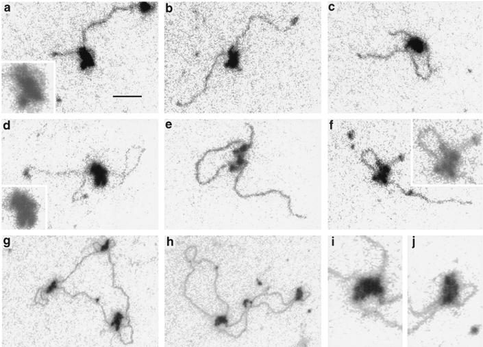
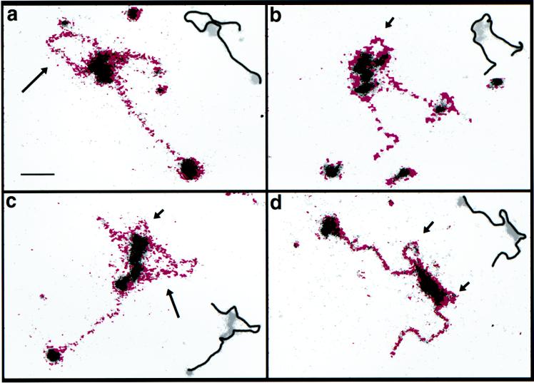
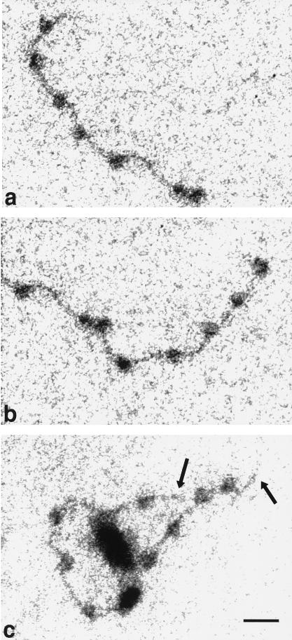
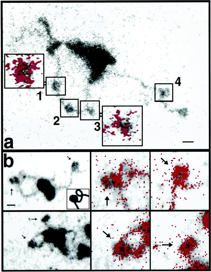
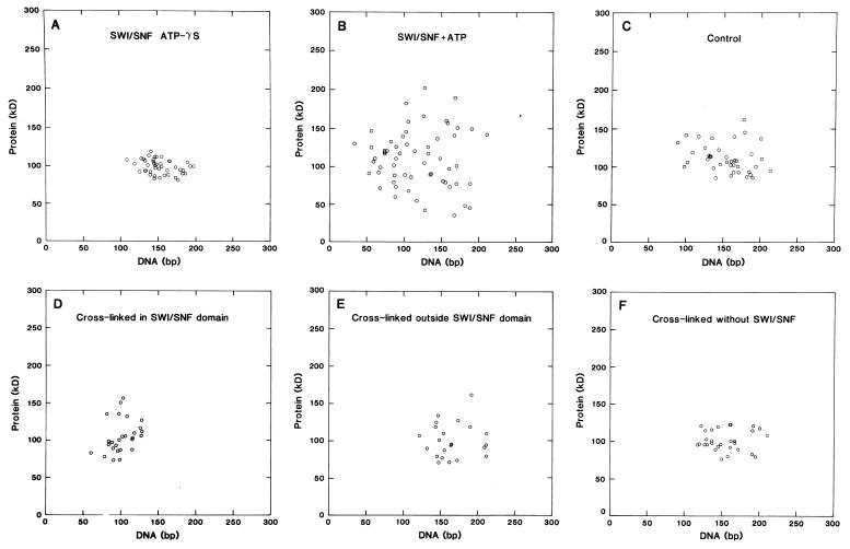
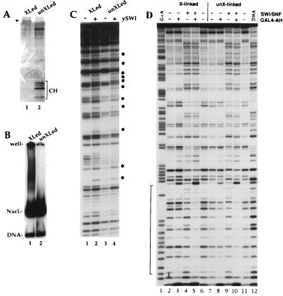
To understand the mechanisms by which the chromatin-remodeling SWI/SNF complex interacts with DNA and alters nucleosome organization, we have imaged the SWI/SNF complex with both naked DNA and nucleosomal arrays by using energy-filtered microscopy. By making ATP-independent contacts with DNA at multiple sites on its surface, SWI/SNF creates loops, bringing otherwise-distant sites into close proximity. In the presence of ATP, SWI/SNF action leads to the disruption of nucleosomes within domains that appear to be topologically constrained by the complex. The data indicate that the action of one SWI/SNF complex on an array of nucleosomes can lead to the formation of a region where multiple nucleosomes are disrupted. Importantly, nucleosome disruption by SWI/SNF results in a loss of DNA content from the nucleosomes. This indicates a mechanism by which SWI/SNF unwraps part of the nucleosomal DNA.


Figures

FIG. 1
FIG. 1
Electron spectroscopic images of SWI/SNF complexes on linear plasmid DNA fragments (a to f) and relaxed closed circular DNA (g to j). (a to c and f) A 1.0-kb fragment from pBluescript; (d and e) 1.4-kb fragment containing promoter sequences of the human proenkephalin gene. The insets in panels a, d, and f are magnified twofold and reproduced at lower contrast to emphasize the subunit structure of SWI/SNF complexes. Panels i and j are magnified regions from panel h. Bar, 53 (a to f) and 160 (g and h) nm.
FIG. 2
FIG. 2
Electron spectroscopic images of SWI/SNF-DNA complexes. A mass-sensitive image is shown in grey levels, on which is superimposed the net phosphorus image in magenta. The short and long arrows indicate small and large loops, respectively, that have been created by SWI/SNF-DNA contacts. The insets reveal the most likely path of the DNA through or around the SWI/SNF-DNA complex, based on the phosphorus map, the contour length, and the topological consistency. Bar, 35 nm.
FIG. 3
FIG. 3
Electron spectroscopic images of a polynucleosome strand in the absence of SWI/SNF (a and b) and a polynucleosome strand with one SWI/SNF complex (c). The left and right halves of the same strand are shown in panels a and b, respectively. The polynucleosome arrays on a 12-mer repeat of the 208-bp sea urchin 5S rRNA gene nucleosome-positioning sequence (25) were formed by salt dialysis with histones purified from chicken erythrocytes (10). The final buffer contained 25 mM HEPES. All of the strands shown were subjected to incubation in buffer containing ATP. The images were recorded at an energy loss of 155 eV. Bar, 24 nm.
FIG. 4
FIG. 4
Electron spectroscopic images of SWI/SNF-polynucleosome strands with histones not chemically cross-linked (a) and with cross-linked histone octamers (b). The mass and phosphorus contents of SWI/SNF-DNA and nucleosome complexes were determined as described previously (3). (a) Protein and DNA contents of the nucleosomes in the numbered boxes are presented in the text. The colored insets show the net phosphorus distributions of the indicated nucleosomes. Bar, 24 nm (insets, 12 nm). (b) Nucleosomes indicated by arrows in the low-magnification views on the left are shown on the right at higher magnification, with the phosphorus represented as red. The inset in the upper-left panel is included to clarify that a protein-free DNA writhe lies just to the right of the nucleosome at the left of the field. Bar, 11 (left) and 5.5 (right) nm.
FIG. 5
FIG. 5
(A and B) Scatter plots of protein content versus DNA content of particles on polynucleosome strands bound by one SWI/SNF complex. Reactions were carried out in the presence of 2 mM ATP-γS (A) or 2 mM ATP (B). (C) Values for particles in polynucleosome strands exposed to ATP but not SWI/SNF in a mock reaction. The mean protein and DNA contents (± SD) are 100 ± 9 kDa and 151 ± 21 bp (n = 42) (A), 111 ± 45 kDa and 118 ± 43 bp (n = 59) (B), and 112 ± 22 kDa and 157 ± 34 bp (n = 40) (C). (D to F) Scatter plots of nucleosome arrays reconstituted with cross-linked histones. Nucleosomes within a loop created by a SWI/SNF complex (D), nucleosomes outside a loop or on strands not bound by SWI/SNF (E), and nucleosomes on arrays not exposed to SWI/SNF (mock-reacted with ATP) (F) were used to obtain the values for these plots. The average amounts of protein and DNA are 101 ± 26 kDa and 101 ± 17 bp (D), 101 ± 24 kDa and 166 ± 27 bp (E), and 102 ± 15 kDa and 155 ± 27 bp (F).
FIG. 6
FIG. 6
Histone octamer cross-linking does not affect nucleosomal DNA perturbation and enhanced GAL4-AH binding by SWI/SNF. Histone cross-linking conditions with dimethyl suberimidate and nucleosome reconstitution by transfer were described previously (31). (A) Sodium dodecyl sulfate-18% polyacrylamide gel showing that cross-linking conditions with dimethyl suberimidate create a protein band at around 100 kDa (arrowhead shows migration of 97.5-kDa marker) with no detectable free histone or partially cross-linked product (50 ng of histone loaded; 172-bp 5S DNA probe [21] used in the reconstitution). Note that the cross-linked histones take up less stain than control histones. The band that migrates more slowly than the core histones corresponds to bovine serum albumin, used to maintain stability. (B) Mobility shift gel showing that the cross-linked octamers do not significantly change the migration of the 5S mononucleosome. (C) Nucleosomes (25 nM) from the preparation used for panel B were incubated with 1 mM ATP in the presence (+) or absence (−) of purified SWI/SNF (2.5 nM) and analyzed by DNase I digestion. The bullets show where the intensity of a band is modified by SWI/SNF action. (D) A single-GAL4-site DNA probe (7) was reconstituted in nucleosome cores by using cross-linked (lanes 2 to 6) or non-cross-linked (lanes 7 to 11) histone octamers. The nucleosomes (25 nM) were incubated in the presence (+) or absence (−) of 15 nM SWI/SNF and/or 100 nM GAL4-AH dimers as indicated and were analyzed by DNase I digestion. Mg-ATP (1 mM) is also present in all of the lanes. The bracket indicates the GAL4-AH binding site. XLed and X-linked, cross-linked; unXLed and unX-linked, not cross-linked.

References

    1. Bazett-Jones D P. Empirical basis for phosphorus mapping and structure determination of DNA: protein complexes by electron spectroscopic imaging. Microbeam Anal. 1993;2:69–79.
    1. Bazett-Jones D P, Leblanc B, Herfort M, Moss T. Short-range DNA looping by the Xenopus HMG-box transcription factor, xUBF. Science. 1994;264:1134–1137. - PubMed
    1. Bazett-Jones D P, Mendez E, Czarnota G J, Ottensmeyer F P, Allfrey V G. Visualization and analysis of unfolded nucleosomes associated with transcribing chromatin. Nucleic Acids Res. 1996;24:321–329. - PMC - PubMed
    1. Cairns B R, Lorch Y, Li Y, Zhang M, Lacomis L, Erdjument-Bromage H, Tempst P, Du J, Laurent B, Kornberg R D. RSC, an essential, abundant chromatin re-modeling complex. Cell. 1996;87:1249–1260. - PubMed
    1. Carlson M, Laurent B C. The SWI/SNF family of global transcriptional activators. Curr Opin Cell Biol. 1994;6:396–402. - PubMed

Publication types

MeSH terms

LinkOut - more resources