. 1999 Feb;19(2):1498-507.
doi: 10.1128/MCB.19.2.1498.

rlk/TXK encodes two forms of a novel cysteine string tyrosine kinase activated by Src family kinases

Affiliations

rlk/TXK encodes two forms of a novel cysteine string tyrosine kinase activated by Src family kinases

J Debnath et al. Mol Cell Biol. 1999 Feb.

Abstract

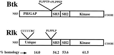
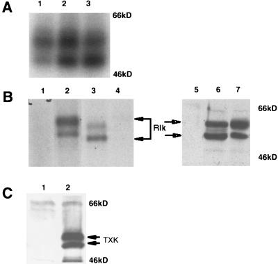
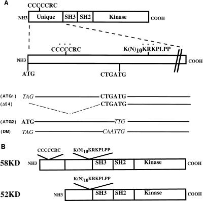
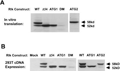
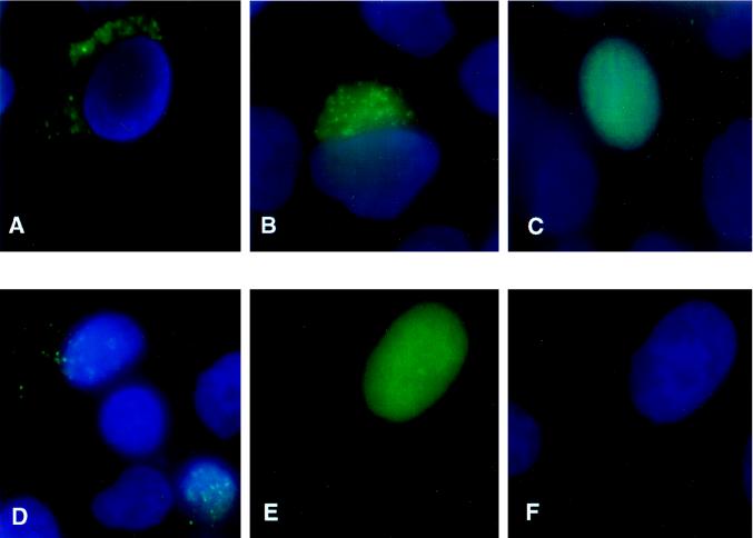
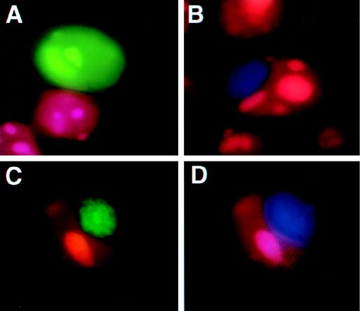
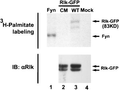
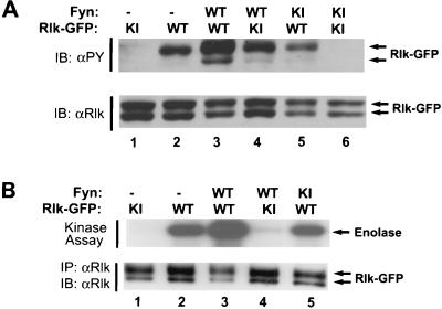
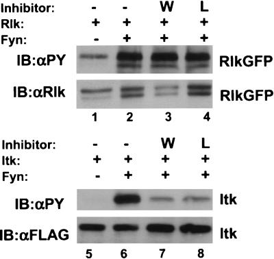
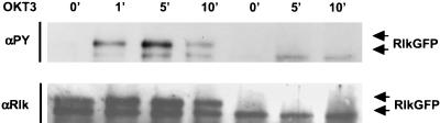
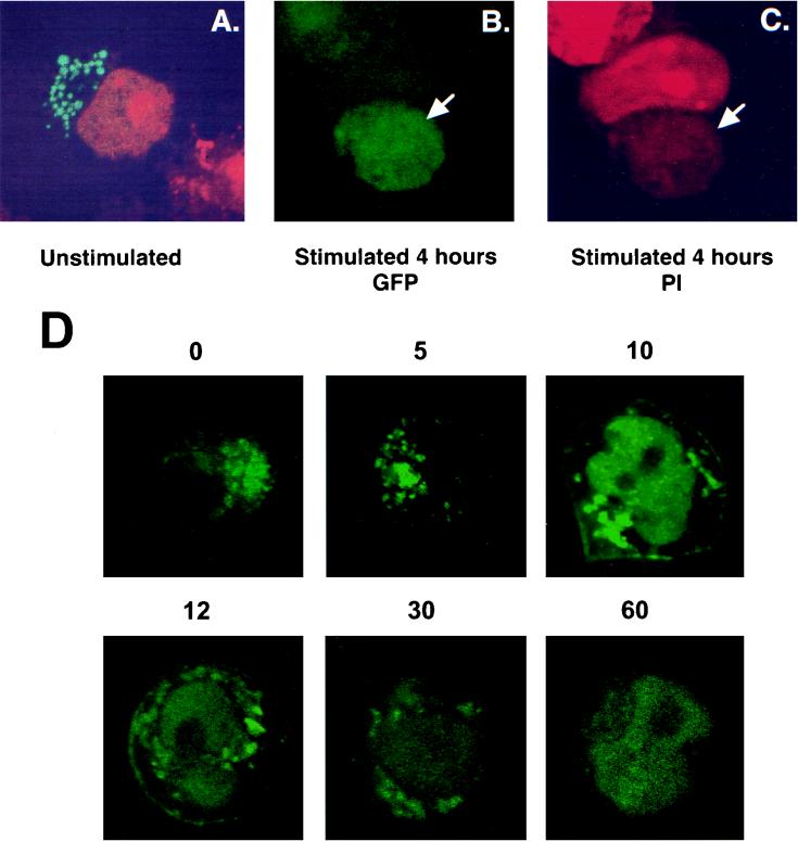
Rlk/Txk is a member of the BTK/Tec family of tyrosine kinases and is primarily expressed in T lymphocytes. Unlike other members of this kinase family, Rlk lacks a pleckstrin homology (PH) domain near the amino terminus and instead contains a distinctive cysteine string motif. We demonstrate here that Rlk protein consists of two isoforms that arise by alternative initiation of translation from the same cDNA. The shorter, internally initiated protein species lacks the cysteine string motif and is located in the nucleus when expressed in the absence of the larger form. In contrast, the larger form is cytoplasmic. We show that the larger form is palmitoylated and that mutation of its cysteine string motif both abolishes palmitoylation and allows the protein to migrate to the nucleus. The cysteine string, therefore, is a critical determinant of both fatty acid modification and protein localization for the larger isoform of Rlk, suggesting that Rlk regulation is distinct from the other Btk family kinases. We further show that Rlk is phosphorylated and changes localization in response to T-cell-receptor (TCR) activation and, like the other Btk family kinases, can be phosphorylated and activated by Src family kinases. However, unlike the other Btk family members, Rlk is activated independently of the activity of phosphatidylinositol 3-kinase, consistent with its lack of a PH domain. Thus, Rlk has two distinct isoforms, each of which may have unique properties in signaling downstream from the TCR.


Figures

FIG. 1
FIG. 1
Comparison of the predicted organizations of Rlk and Btk. Percent amino acid identities are indicated at the bottom of the figure.
FIG. 2
FIG. 2
Rlk encodes two proteins. (A) In vitro protein kinase assay of Rlk protein immunoprecipitated with anti-Rlk antiserum from lysates of murine lymph node cells (lane 1), splenocytes (lane 2), or thymocytes (lane 3). Immune complexes were labeled with [γ-32P]ATP, resolved by SDS-PAGE, and visualized by autoradiography. (B) (Lanes 1 to 4) In vitro protein kinase assay of Rlk protein immunoprecipitated with anti-Rlk antiserum from lysates of 293T cells transfected with vector alone (lane 1) or murine myc-tagged rlk cDNA (lane 2) compared with immunoprecipitates from murine thymocytes (lane 3). Lane 4 contains immunoprecipitates with preimmune sera from thymocyte lysates. The two major protein species generated from the Rlk-myc construct migrate slightly slower than endogenous Rlk in T cells, as expected from the 15-amino-acid tag (lanes 3 and 2). Versions of the rlk cDNA lacking the myc insert generated proteins that comigrated with the endogenous forms (data not shown). (Lanes 5 to 7) Immunoblot analysis with anti-Rlk of 293T cell lysates from cells that were mock transfected (lane 5) or transfected with murine rlk cDNA (lane 6) compared to in vitro transcription-translation products (lane 7) generated from rlk cDNA. (C) Immunoblot analysis with anti-human TXK antisera of 293T cell lysates from cells that were mock transfected (lane 1) or transfected with human Txk cDNA (lane 2). The expected TXK products are indicated.
FIG. 3
FIG. 3
Schematic of Rlk mutants. (A) Rlk protein organization and locations of the cysteine string motif and putative NLS in the amino terminus of Rlk. Residues changed in the mutants of the cysteine string motif and NLS are marked by asterisks. The relative positions of the putative translational initiation sites, as well as the nucleotide sequences of the various start site mutant derivatives (italicized), are shown below the schematic. SH3, Src homology 3; SH2, Src homology 2. (B) Schematic organization of the two forms of Rlk.
FIG. 4
FIG. 4
Distinct protein isoforms of rlk are generated by the utilization of alternative translational start codons. (A) In vitro transcription-translation products generated from WT and mutant rlk cDNAs, resolved by SDS-PAGE, and visualized by fluorography. (B) Expression of rlk WT and mutant cDNAs in 293T cells. Equal amounts of protein from total cellular lysates of cells transfected with the indicated constructs were resolved by SDS-PAGE and visualized by immunoblotting with anti-Rlk serum (lower). Mutants are described in Fig. 3.
FIG. 5
FIG. 5
Subcellular localization of the two forms of WT and mutant Rlk-GFP fusions. HeLa cells were transiently transfected with the following constructs, fixed, and stained with DAPI. (A) WT GFP-Rlk. (B) AUG2Rlk-GFP expressing only the long form. (C) AUG1Rlk-GFP expressing only the short form. (D) NLS mutant of the short form of Rlk-GFP (NLS-AUG1Rlk-GFP). (E and F) Cysteine mutant of the long form of Rlk (CMRlk-GFP) with fluorescein channel only (E) or DAPI only (F).
FIG. 6
FIG. 6
The two forms of Rlk colocalize when coexpressed. 293T cells were transiently transfected with Rlk-GFP expression constructs, fixed, and counterstained with propidium iodide. (A) AUG1Rlk-GFP (short form). (B) AUG2Rlk-BFP (long form). (C and D) AUG2Rlk-BFP (long form) and AUG1Rlk-GFP (short form) cotransfected and visualized for GFP (C) or visualized for BFP (D). The colocalizations of the two forms of Rlk are indicated by arrows.
FIG. 7
FIG. 7
Subcellular fractionation of murine thymocytes. Cells were fractionated as described in the text, and the nuclear and cytoplasmic fractions from 6 × 10⁷ cells were immunoprecipitated and analyzed by kinase assay. Nucl, nuclear pellet; Cyto, S100 supernatant.
FIG. 8
FIG. 8
WT Rlk is modified by palmitoylation. Mutation of the cysteine string abolishes [3H]palmitate labeling. 293T cells were transfected with cDNAs encoding Fyn (positive control, lane 1), a cysteine mutant of Rlk-GFP (lane 2), WT Rlk (lane 3), or control vector alone (lane 4). Twenty-four hours after transfection, cells were labeled with [3H]palmitate for 2 h, washed, and lysed, and Rlk or Fyn was immunoprecipitated as described in the text. (Top) Samples were analyzed by SDS–10% PAGE and visualized by fluorography. (Bottom) Immunoblot (IB) for Rlk.
FIG. 9
FIG. 9
Tyrosine phosphorylation and activation of Rlk kinase upon coexpression with Fyn in 293T cells. (A) 293T cells were transfected with Rlk-GFP fusion and FynT expression constructs, followed by cell lysis at 24 h. Equal amounts of detergent-soluble lysates were resolved by SDS-PAGE and subjected to antiphosphotyrosine (4G10) (top) or anti-Rlk (bottom) immunoblotting (IB). (B) Immune complex kinase assays. Cells expressing Rlk-GFP fusion proteins alone or with Fyn were lysed and immunoprecipitated with anti-Rlk. Immune complexes were washed and subjected to an in vitro kinase assay with [γ-32P]ATP and acid-denatured enolase as an exogenous substrate. Phosphorylated proteins were resolved by SDS-PAGE and detected by autoradiography. (Top) In vitro kinase activity on enolase. (Bottom) anti-Rlk. Equivalent expression of Fyn was confirmed by immunoblotting with anti-Fyn (data not shown). WT, wild type (kinase active); KI, kinase inactive.
FIG. 10
FIG. 10
Rlk activation by Fyn is not affected by inhibitors of PI 3-kinase. (Top) Cells were transfected with constructs expressing either Rlk alone or Rlk plus Fyn. Twenty-four hours after transfection, cells were split equally and treated 24 h later with either dimethyl sulfoxide (DMSO) (lanes 1 and 2), 1 mM Wortmannin (lane 3), or 100 mM Ly294002 (lane 4). After 30 min, cells were lysed, resolved by SDS-PAGE, and examined by Western blotting with antiphosphotyrosine (top row) or anti-Rlk (bottom row). (Bottom) Cells were transfected with constructs expressing either Itk-FLAG alone or Itk-FLAG plus Fyn and treated as above with either DMSO (lanes 5 and 6), 1 mM Wortmannin (lane 7), or 100 mM Ly294002 (lane 8). Western blotting was performed with antiphosphotyrosine (top row) or anti-FLAG (bottom row).
FIG. 11
FIG. 11
Tyrosine phosphorylation of Rlk-GFP in Jurkat cells. Cells were electroporated with either WT Rlk-GFP or AUG2Rlk-GFP expressing only the short form of Rlk. Twenty-four hours later, cells were stimulated with anti-CD3 antibody OKT3 for the indicated times (in minutes). Cells were lysed and Rlk was immunoprecipitated. Immunoprecipitated Rlk was immunoblotted with antiphosphotyrosine (4G10) (top) or anti-Rlk (bottom).
FIG. 12
FIG. 12
Activation of Jurkat cells changes the subcellular localization of Rlk. Cells were electroporated with WT Rlk-GFP and stimulated 24 h later with either OKT3 or PMA and ionomycin, counterstained with propidium iodide, and examined by confocal microscopy. (A) Unstimulated cells. (B) Stimulated cells, fluorescein channel. (C) Same as panel B, with rhodamine channel for visualizing propidium iodide. (D) Time course of localization of Rlk-GFP after TCR stimulation. Cells were visualized live at the indicated times (in minutes) after treatment with anti-CD3.

References

    1. August A, Gibson S, Kawakami Y, Kawakami T, Mills G B, Dupont B. CD28 is associated with and induces the immediate tyrosine phosphorylation and activation of the Tec family kinase ITK/EMT in the human Jurkat leukemic T-cell line. Proc Natl Acad Sci USA. 1994;91:9347–9351. - PMC - PubMed
    1. August A, Sadra A, Dupont B, Hanafusa H. Src-induced activation of inducible T cell kinase (ITK) requires phosphatidylinositol 3-kinase activity and the Pleckstrin homology domain of inducible T cell kinase. Proc Natl Acad Sci USA. 1997;94:11227–11232. - PMC - PubMed
    1. Braun J E, Scheller R H. Cysteine string protein, a DnaJ family member, is present on diverse secretory vesicles. Neuropharmacology. 1995;34:1361–1369. - PubMed
    1. Cheng, G., M. Chamorro, and P. L. Schwartzberg. Unpublished observations.
    1. Debnath, J. Unpublished observations.

MeSH terms

LinkOut - more resources