. 1999 Feb;19(2):1569-81.
doi: 10.1128/MCB.19.2.1569.

The MKK7 gene encodes a group of c-Jun NH2-terminal kinase kinases

Affiliations

The MKK7 gene encodes a group of c-Jun NH2-terminal kinase kinases

C Tournier et al. Mol Cell Biol. 1999 Feb.

Abstract

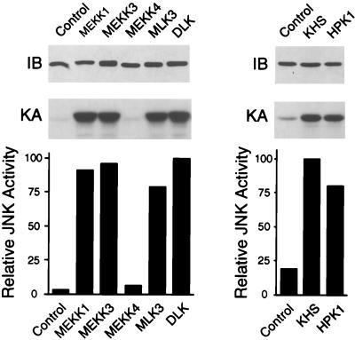
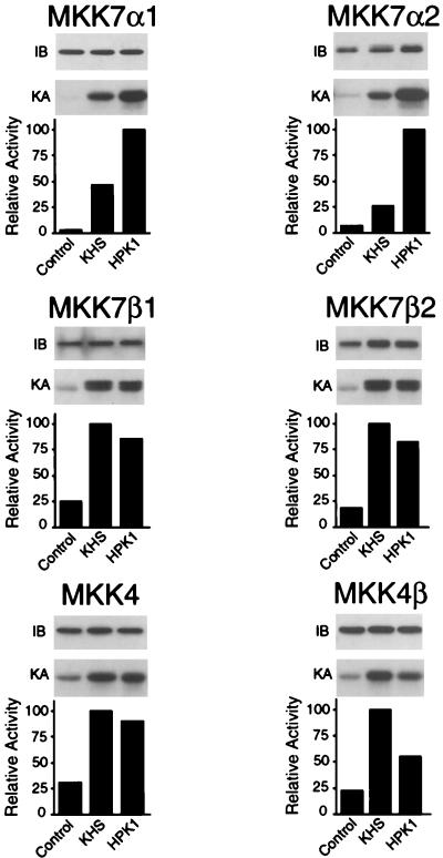
The c-Jun NH2-terminal protein kinase (JNK) is a member of the mitogen-activated protein kinase (MAPK) group and is an essential component of a signaling cascade that is activated by exposure of cells to environmental stress. JNK activation is regulated by phosphorylation on both Thr and Tyr residues by a dual-specificity MAPK kinase (MAPKK). Two MAPKKs, MKK4 and MKK7, have been identified as JNK activators. Genetic studies demonstrate that MKK4 and MKK7 serve nonredundant functions as activators of JNK in vivo. We report here the molecular cloning of the gene that encodes MKK7 and demonstrate that six isoforms are created by alternative splicing to generate a group of protein kinases with three different NH2 termini (alpha, beta, and gamma isoforms) and two different COOH termini (1 and 2 isoforms). The MKK7alpha isoforms lack an NH2-terminal extension that is present in the other MKK7 isoforms. This NH2-terminal extension binds directly to the MKK7 substrate JNK. Comparison of the activities of the MKK7 isoforms demonstrates that the MKK7alpha isoforms exhibit lower activity, but a higher level of inducible fold activation, than the corresponding MKK7beta and MKK7gamma isoforms. Immunofluorescence analysis demonstrates that these MKK7 isoforms are detected in both cytoplasmic and nuclear compartments of cultured cells. The presence of MKK7 in the nucleus was not, however, required for JNK activation in vivo. These data establish that the MKK4 and MKK7 genes encode a group of protein kinases with different biochemical properties that mediate activation of JNK in response to extracellular stimuli.


Figures

FIG. 1
FIG. 1
Primary structures of MKK7 protein kinase isoforms. The primary sequence of the mouse MKK7 protein kinase isoforms deduced from the sequences of cDNA clones are presented in single-letter code. Residues that are identical to those in MKK7γ2 (.), deletions (−), and termination codons (#) are indicated.
FIG. 2
FIG. 2
The MKK7 gene is located on mouse chromosome 11. The MKK7 gene was identified by FISH analysis of murine metaphase chromosomes by using an MKK7 genomic clone as a probe. The FISH signal is illustrated on the left (arrow). The corresponding DAPI signal indicating chromosome 11 is shown on the right. Detailed comparison of DAPI-banded chromosome 11 with the FISH signal indicated that the MKK7 gene is located in region A2.
FIG. 3
FIG. 3
Schematic representation of the structure of the MKK7 gene. The MKK7 gene is formed by 14 exons. The alternative splicing that creates the α, β, γ, 1, and 2 isoforms of MKK7 is illustrated. The coding (black) and noncoding (grey) regions and excluded exons (striped) are shown. Initiation codons (ATG), termination codons (∗), and polyadenylation signals (•) are indicated.
FIG. 4
FIG. 4
Effects of MKK7 homologs on JNK activation. (A) COS-7 cells were transfected with expression vectors encoding Flag epitope-tagged MKK7α1, MKK7α2, MKK7β1, MKK7β2, MKK7γ1, or MKK7γ2. The expression of each of these MKK7 isoforms was examined by immunoblot (IB) analysis using the Flag-specific monoclonal antibody M2. The MKK7 isoforms were immunoprecipitated, and their activities were measured in the immune complex by a coupled protein kinase assay (KA) using recombinant GST-JNK1 and GST–c-Jun as substrates. The phosphorylated c-Jun was detected after SDS-PAGE by autoradiography and was quantitated by PhosphorImager analysis. The effect of cotransfection with an empty expression vector (−) or with an MEKK1 expression vector (+) is shown. Similar results were observed in three separate experiments. (B) MKK7 activity, presented as relative protein kinase activity. (C) MKK7 activation caused by MEKK1 (fold activity over MKK7 activity in the absence of MEKK1).
FIG. 5
FIG. 5
Interaction of JNK with MKK7 isoforms. (A) Selective binding of MKK7β isoforms to JNK. Epitope-tagged JNK1 and either GST (Control) or GST-tagged MKK7α1, MKK7α2, MKK7β1, or MKK7β2 were expressed in COS-1 cells. Protein expression levels, monitored by immunoblot analysis of the cell lysates, were similar in all transfections. The MKK7 protein kinases were isolated from cell extracts by incubation with GSH-agarose. The binding of JNK1 was examined by immunoblot analysis with an antibody to the HA epitope tag. (B) JNK binds to the NH2 terminus of MKK7β. Bacterially expressed GST (Control) or a GST fusion protein (residues 1 to 73 of MKK7β) was immobilized on GSH-agarose and incubated with extracts prepared from COS-7 cells expressing epitope-tagged JNK1. The agarose beads were washed, and the amount of bound JNK1 was examined by immunoblot analysis.
FIG. 6
FIG. 6
Effect of MEKK and STE20 protein kinases on JNK activation. To examine the ability of MAPKKKs and STE20 homologs to activate JNK1, COS-7 cells were cotransfected with a mammalian expression vector encoding HA-tagged JNK1 together with an empty expression vector (Control) or an expression vector encoding either MEKK1, MEKK3, MEKK4, MLK3, DLK, KHS, or HPK1. The expression of JNK1 was examined by immunoblot (IB) analysis using a monoclonal antibody to the HA epitope tag. JNK1 was immunoprecipitated and its activity was measured in the immune complex by a protein kinase assay (KA) using recombinant GST–c-Jun as the substrate. The phosphorylated c-Jun was detected after SDS-PAGE by autoradiography and was quantitated by PhosphorImager analysis. JNK activity is presented as relative protein kinase activity. The levels of JNK activation caused by MEKK1, MEKK3, MEKK4, MLK3, DLK, KHS, and HPK1 were 28-, 29-, 2.0-, 24-, 30-, 5.3-, and 4.3-fold, respectively. Similar results were obtained in three separate experiments.
FIG. 7
FIG. 7
Activation of MKK4 and MKK7 protein kinases by MAPKKKs. To examine the abilities of MAPKKKs to activate MKK isoforms, COS-7 cells were cotransfected with mammalian expression vectors encoding tagged MKK7α1, MKK7α2, MKK7β1, MKK7β2, MKK4, or MKK4β together with either an empty expression vector (Control) or an expression vector encoding MEKK1, MEKK3, MEKK4, MLK3, or DLK. MKK protein expression was monitored by immunoblot (IB) analysis. The MKK4 and MKK7 isoforms were immunoprecipitated, and their activities were measured by a coupled protein kinase assay (KA) using recombinant JNK1 and c-Jun as substrates. The phosphorylated c-Jun was detected after SDS-PAGE by autoradiography and was quantitated by PhosphorImager analysis. MAPKK activity is presented as relative protein kinase activity. The levels of MAPKK activation caused by MEKK1, MEKK3, MEKK4, MLK3, and DLK were 27-, 19-, 1.8-, 5.0-, and 7.0-fold (MKK7α1); 32-, 10-, 1.4-, 13-, and 6.5-fold (MKK7α2); 5.6-, 2.0-, 0.3-, 4.2-, and 4.5-fold (MKK7β1); 13-, 2.6-, 1.3-, 7.5-, and 5.1-fold (MKK7β2); 42-, 88-, 2.0-, 38-, and 39-fold (MKK4); and 53-, 77-, 2.2-, 48-, and 36-fold (MKK4β), respectively. Similar results were obtained in three separate experiments.
FIG. 8
FIG. 8
Activation of MKK4 and MKK7 protein kinases by STE20 homologs. To examine the abilities of KHS and HPK1 to activate MKK7 isoforms, COS-7 cells were cotransfected with Flag-tagged MKK7α1, MKK7α2, MKK7β1, MKK7β2, MKK4, or MKK4β together with an empty expression vector (Control) or an expression vector for either KHS or HPK1. Protein expression was monitored by immunoblot (IB) analysis. Flag-tagged MKKs were immunoprecipitated, and MKK activity was measured in the immune complex by a coupled protein kinase assay (KA) using recombinant JNK1 and c-Jun as substrates. The phosphorylated c-Jun was detected after SDS-PAGE by autoradiography and quantitated by PhosphorImager analysis. MAPKK activity is presented as relative protein kinase activity. The levels of MAPKK activation caused by KHS and HPK were 15- and 31-fold (MKK7α1); 3.9- and 15-fold (MKK7α2); 4.0- and 3.5-fold (MKK7β1); 5.4- and 4.4-fold (MKK7β2); 3.2- and 2.9-fold (MKK4); and 4.4- and 2.4-fold (MKK4β), respectively. Similar results were obtained in three separate experiments.
FIG. 9
FIG. 9
Regulation of MKK4 and MKK7 protein kinases by extracellular stimuli. 293 cells were transiently transfected with expression vectors (pEBG) encoding either MKK7α1, MKK7α2, MKK7β1, MKK7β2, MKK4, or MKK4β; 36 h after transfection, the cells were untreated (Control) or treated with UV-C (UV; 80 J/m2), anisomycin (ANISO; 5 μg/ml), TNF-α (20 ng/ml), or IL-1α (2 ng/ml). The cells were harvested after incubation at 37°C (30 min). MKK4 and MKK7 were isolated by using GSH-Sepharose beads, and the MKK activity was measured by a coupled protein kinase assay using recombinant JNK1 and c-Jun as substrates. The radioactivity incorporated into c-Jun was quantitated after SDS-PAGE by PhosphorImager analysis. MAPKK activity is presented as relative protein kinase activity. The levels of MAPKK activation caused by UV, anisomycin, TNF-α, and IL-1α were 1.4-, 1.5-, 2.2-, and 1.9-fold (MKK7α1); 2.8-, 1.8-, 3.9-, and 2.5-fold (MKK7α2); 2.4-, 2.1-, 2.5-, and 4.1-fold (MKK7β1); 2.0-, 1.7-, 2.8-, and 2.9-fold (MKK7β2); 4.7-, 3.2-, 0.9-, and 0.9-fold (MKK4); and 2.6-, 2.3-, 0.9-, and 0.9-fold (MKK4β), respectively. Similar results were obtained in three separate experiments.
FIG. 10
FIG. 10
Subcellular distribution of MAPKK isoforms. (A) Endogenous MKK4 and MKK7 (red) were detected by immunofluorescence analysis using primary antibodies specific for these MKK isoforms. The cells were untreated (Control) or treated (30 min) with UV (80 J/m2) or IL-1α (10 ng/ml). DNA was visualized by staining with DAPI (blue). The scale bar (white) represents 20 μm. (B) Epitope-tagged MKK7α1, MKK7α2, MKK7β1, MKK7β2, MKK4, MKK4β, MEK1, and ΔN3-MEK1-EE were detected by using a primary monoclonal antibody to the epitope tag and a Texas red-labeled secondary antibody. The scale bar (white) represents 10 μm.
FIG. 11
FIG. 11
Effect of an NES on the properties of MKK7. (A) Schematic representation of MKK7β and NES-MKK7β. Both proteins were constructed with an NH2-terminal Flag epitope (grey box). The nuclear export signal (NES) of MEK1 (residues 32 to 51) was inserted following the Flag epitope to create NES-MKK7β. (B) The MKK7 proteins and HA-JNK1 were expressed in COS-7 cells. Transfected cells were identified by immunofluorescence analysis using antibody M2, which binds the Flag epitope on the MKK7 proteins. Activated JNK in the transfected cells was examined by using an antibody to phospho-JNK [JNK(P)]. The MKK7 (red) and phospho-JNK (green) were detected with secondary antibodies conjugated to Texas red and fluorescein, respectively. DNA was visualized by staining with DAPI (blue). The scale bar (white) represents 10 μm. (C) Regulation of MKK7β2 and NES-MKK7β2 by extracellular stimuli. COS-7 cells expressing MKK7β2 or NES-MKK7β2 were untreated (Control) or treated (30 min) with UV-C (80 J/m2) or IL-1α (10 ng/ml). The expression of MKK7 was examined by immunoblot (IB) analysis using the Flag-specific monoclonal antibody M2. MKK7 activity was measured in a coupled protein kinase assay (KA). The radioactivity incorporated into c-Jun was quantitated after SDS-PAGE by PhosphorImager analysis. MAPKK activity is presented as relative protein kinase activity. The levels of MAPKK activation caused by UV and IL-1α were 3.0- and 3.7-fold (MKK7β2) and 1.5- and 1.8-fold (NES-MKK7β2), respectively. (D) Activation of JNK1 by MKK7β and NES-MKK7β. COS-7 cells were cotransfected with plasmids expressing HA-tagged JNK1 together with an empty vector (Control) or an expression vector encoding Flag-tagged MKK7β1, NES-MKK7β1, MKK7β2, or NES-MKK7β2. The expression of MKK7 and JNK1 was monitored by immunoblot (IB) analysis using antibodies to Flag and HA, respectively. JNK1 activity was measured by immune complex kinase assay (KA) using the substrate c-Jun. The radioactivity incorporated into c-Jun was quantitated after SDS-PAGE by PhosphorImager analysis. JNK activity is presented as relative protein kinase activity. The levels of JNK activation observed for MKK7β1, NES-MKK7β1, MKK7β2, and NES-MKK7β2 were 21-, 27-, 25-, and 31-fold, respectively.
FIG. 11
FIG. 11
Effect of an NES on the properties of MKK7. (A) Schematic representation of MKK7β and NES-MKK7β. Both proteins were constructed with an NH2-terminal Flag epitope (grey box). The nuclear export signal (NES) of MEK1 (residues 32 to 51) was inserted following the Flag epitope to create NES-MKK7β. (B) The MKK7 proteins and HA-JNK1 were expressed in COS-7 cells. Transfected cells were identified by immunofluorescence analysis using antibody M2, which binds the Flag epitope on the MKK7 proteins. Activated JNK in the transfected cells was examined by using an antibody to phospho-JNK [JNK(P)]. The MKK7 (red) and phospho-JNK (green) were detected with secondary antibodies conjugated to Texas red and fluorescein, respectively. DNA was visualized by staining with DAPI (blue). The scale bar (white) represents 10 μm. (C) Regulation of MKK7β2 and NES-MKK7β2 by extracellular stimuli. COS-7 cells expressing MKK7β2 or NES-MKK7β2 were untreated (Control) or treated (30 min) with UV-C (80 J/m2) or IL-1α (10 ng/ml). The expression of MKK7 was examined by immunoblot (IB) analysis using the Flag-specific monoclonal antibody M2. MKK7 activity was measured in a coupled protein kinase assay (KA). The radioactivity incorporated into c-Jun was quantitated after SDS-PAGE by PhosphorImager analysis. MAPKK activity is presented as relative protein kinase activity. The levels of MAPKK activation caused by UV and IL-1α were 3.0- and 3.7-fold (MKK7β2) and 1.5- and 1.8-fold (NES-MKK7β2), respectively. (D) Activation of JNK1 by MKK7β and NES-MKK7β. COS-7 cells were cotransfected with plasmids expressing HA-tagged JNK1 together with an empty vector (Control) or an expression vector encoding Flag-tagged MKK7β1, NES-MKK7β1, MKK7β2, or NES-MKK7β2. The expression of MKK7 and JNK1 was monitored by immunoblot (IB) analysis using antibodies to Flag and HA, respectively. JNK1 activity was measured by immune complex kinase assay (KA) using the substrate c-Jun. The radioactivity incorporated into c-Jun was quantitated after SDS-PAGE by PhosphorImager analysis. JNK activity is presented as relative protein kinase activity. The levels of JNK activation observed for MKK7β1, NES-MKK7β1, MKK7β2, and NES-MKK7β2 were 21-, 27-, 25-, and 31-fold, respectively.

Similar articles

Cited by

References

    1. Ahn N G, Seger R, Krebs E G. The mitogen-activated protein kinase activator. Curr Opin Cell Biol. 1992;4:992–999. - PubMed
    1. Anafi M, Kiefer F, Gish G D, Mbamalu G, Iscove N N, Pawson T. SH2/SH3 adaptor proteins can link tyrosine kinases to a Ste20-related protein kinase, HPK1. J Biol Chem. 1997;272:27804–27811. - PubMed
    1. Bagrodia S, Derijard B, Davis R J, Cerione R A. Cdc42 and PAK-mediated signaling leads to Jun kinase and p38 mitogen-activated protein kinase activation. J Biol Chem. 1995;270:27995–27998. - PubMed
    1. Bardwell L, Thorner J. A conserved motif at the amino termini of MEKs might mediate high-affinity interaction with the cognate MAPKs. Trends Biochem Sci. 1996;21:373–374. - PubMed
    1. Ben-Levy R, Hooper S, Wilson R, Paterson H F, Marshall C J. Nuclear export of the stress-activated protein kinase p38 mediated by its substrate MAPKAP kinase-2. Curr Biol. 1998;8:1049–1057. - PubMed

Publication types

MeSH terms