. 1999 Feb;19(2):1582-94.
doi: 10.1128/MCB.19.2.1582.

Thymidylate synthase protein and p53 mRNA form an in vivo ribonucleoprotein complex

Affiliations

Thymidylate synthase protein and p53 mRNA form an in vivo ribonucleoprotein complex

E Chu et al. Mol Cell Biol. 1999 Feb.

Abstract

A thymidylate synthase (TS)-ribonucleoprotein (RNP) complex composed of TS protein and the mRNA of the tumor suppressor gene p53 was isolated from cultured human colon cancer cells. RNA gel shift assays confirmed a specific interaction between TS protein and the protein-coding region of p53 mRNA, and in vitro translation studies demonstrated that this interaction resulted in the specific repression of p53 mRNA translation. To demonstrate the potential biological role of the TS protein-p53 mRNA interaction, Western immunoblot analysis revealed nearly undetectable levels of p53 protein in TS-overexpressing human colon cancer H630-R10 and rat hepatoma H35(F/F) cell lines compared to the levels in their respective parent H630 and H35 cell lines. Polysome analysis revealed that the p53 mRNA was associated with higher-molecular-weight polysomes in H35 cells compared to H35(F/F) cells. While the level of p53 mRNA expression was identical in parent and TS-overexpressing cell lines, the level of p53 RNA bound to TS in the form of RNP complexes was significantly higher in TS-overexpressing cells. The effect of TS on p53 expression was also investigated with human colon cancer RKO cells by use of a tetracycline-inducible system. Treatment of RKO cells with a tetracycline derivative, doxycycline, resulted in 15-fold-induced expression of TS protein and nearly complete suppression of p53 protein expression. However, p53 mRNA levels were identical in transfected RKO cells in the absence and presence of doxycycline. Taken together, these findings suggest that TS regulates the expression of p53 at the translational level. This study identifies a novel pathway for regulating p53 gene expression and expands current understanding of the potential role of TS as a regulator of cellular gene expression.


Figures

FIG. 1
FIG. 1
(A) Specific interaction between human TS and p53 RNA. RNA EMSAs were performed with a 1,587-nt radiolabeled p53 RNA probe (2.2 fmol, 100,000 cpm) incubated in the absence (lane 1) or presence of partially purified human recombinant TS (specific activity, 0.01 U/mg; 3 pmol; lane 2) and pure human recombinant TS (specific activity, 0.7 U/mg; 3 pmol; lane 3). Pure human recombinant TS (3 pmol) was preincubated with proteinase K (1 μg/μl) for 15 min (lane 4) or heat denatured at 95°C for 15 min (lane 5) and then included in a reaction mixture with a radiolabeled p53 RNA probe. Labeled p53 RNA was incubated with pure human recombinant deoxycytidylate deaminase (specific activity, 1,000 U/mg; 3 pmol; lane 6). (B) Effect of nucleotides on RNA binding of TS protein. Radiolabeled p53 RNA (2.2 fmol; 100,000 cpm) was incubated in the absence (lane 1) or presence (lanes 2 to 4) of human recombinant TS (3 pmol). TS protein was preincubated with either dUMP (30 μM; lane 3) or the 5-FU metabolite FdUMP (30 μM; lane 4) for 15 min, followed by the addition of radiolabeled p53 RNA probe. Reactions were performed at room temperature for 15 min. Then, RNase T1 (12 U) was added for 10 min, followed by heparin (5 mg/ml) for an additional 10 min. Samples were electrophoresed in a nondenaturing 4% acrylamide gel. The specific complex is indicated by the arrows.
FIG. 2
FIG. 2
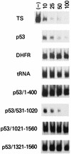
Competition experiment to determine the relative binding of various p53 RNA sequences. Radiolabeled p53 RNA (2.2 fmol; 100,000 cpm) was incubated with TS (3 pmol) alone (−) or in the presence of a 0- to 100-fold molar excess of either the 1,524-nt TS RNA, the 1,587-nt p53 RNA, or the 700-nt DHFR RNA, yeast tRNA, p53/1-400 RNA, p53/531-1020 RNA, p53/1021-1560 RNA, or p53/1321-1560 RNA. Each RNA sequence was synthesized in vitro as described in Materials and Methods.
FIG. 3
FIG. 3
(A) Dose-dependent inhibition of p53 mRNA translation in vitro by TS. Translation reaction mixtures containing rabbit reticulocyte lysate were incubated with either no exogenous RNA (lane 1) or p53 mRNA (0.2 pmol; lanes 2 to 6). Various amounts of TS protein—1 (lane 3), 2 (lane 4), 3 (lane 5), and 4 (lane 6) pmol—were added to the reaction mixture as indicated. (B) Specificity of inhibition of p53 mRNA translation in vitro by TS. Translation reaction mixtures were incubated with either no RNA (lane 1) or p53 mRNA (0.2 pmol; lanes 2 to 7), and TS (3 pmol; lanes 3 to 5) was included in the reaction mixture as indicated. TS was treated with either proteinase K (1 μg/μl) (lane 4) for 15 min or heat denaturation at 95°C (lane 5) for 15 min and then added to p53 mRNA-containing rabbit lysate reaction mixtures. p53 mRNA was incubated with bovine serum albumin (3 pmol; lane 6) or recombinant deoxycytidylate deaminase (3 pmol; lane 7). (C) Translation reaction mixtures were incubated with 0.2 pmol of yeast mRNA (lanes 1 and 2), 0.2 pmol of human DHFR mRNA (lanes 3 and 4), and 0.2 pmol of human chromogranin A mRNA (lanes 5 and 6). TS (3 pmol) was included in the rabbit reticulocyte lysate reaction mixtures as indicated. (D) p53 mRNA (0.2 pmol) was incubated in the absence (lane 1) or presence (lanes 2 to 4) of human TS protein (3 pmol) as indicated. The nucleotides dUMP (30 μM; lane 3) and FdUMP (30 μM; lane 4) were included in the reaction mixtures. (E) TS mRNA (0.2 pmol) was incubated alone (lane 1) or in the presence of TS protein (3 pmol; lanes 2 to 8). Various exogenous RNAs were also included in the rabbit reticulocyte lysate mixtures: p53/531-1020 RNA at 2 pmol (lane 3) and 4 pmol (lane 4), 1,587-nt p53 mRNA at 2 pmol (lane 5) and 4 pmol (lane 6), p53/1-400 RNA (4 pmol; lane 7), and p53/1321-1560 RNA (4 pmol; lane 8). All translation reaction mixtures were incubated at 37°C for 60 min, and protein products were analyzed by SDS-PAGE and autoradiography. The TS and p53 protein products are indicated by the arrows.
FIG. 4
FIG. 4
Specific effect of TS protein on p53 mRNA translation. p53 mRNA (1,587 nts) was incubated in rabbit reticulocyte lysates in the absence (lane 1) or presence (lane 2) of human TS protein (3 pmol). At the end of the reaction, RNA was extracted from each reaction sample and incubated in a fresh rabbit reticulocyte lysate (lanes 3 and 4) at 37°C for an additional 60 min. Protein products were analyzed by SDS-PAGE and autoradiography. The p53 protein product is indicated by the arrow.
FIG. 5
FIG. 5
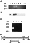
RT-PCR analysis of p53 RNA immunoprecipitated from human colon cancer cells (H630-R10). (A) Ethidium bromide stain. (B) Southern hybridization. The 1,587-nt p53 cDNA was used as a DNA template in a PCR amplification with p53-specific primers p53-3 and p53-4 (lane 1). Whole-cell extracts were immunoprecipitated with either anti-TS polyclonal antibody (lane 3), anti-TS monoclonal antibody (lane 4), no antibody (lane 6), preimmune antisera (lane 7), or anti–alpha-tubulin monoclonal antibody (lane 8) as described in Materials and Methods. The isolated nucleic acid fraction was reverse transcribed and PCR amplified with p53-specific primers. The immunoprecipitated nucleic acid in lane 5 was treated with RNase A prior to RT. A control PCR was performed with p53-specific primers and no DNA template (lane 2). BP, base pairs. (C) Analysis of immunoprecipitated RNAs from human colon cancer cells. RNAs isolated by immunoprecipitation of H630-R10 cell extracts with anti-TS polyclonal antibody were RT-PCR amplified with either p53-specific primers to yield DNA products corresponding to nts 531 to 1020 (lane 1) or max-specific (lane 2), DHFR-specific (lane 3), or GAPDH-specific (lane 4) primer sets. Amplified DNA products were resolved on a 1% nondenaturing agarose gel and analyzed by staining with ethidium bromide. (D) p53 RNA sequence. The p53 RNA sequence predicted to be RT-PCR amplified is shown in relation to its position on the full-length p53 mRNA. The translational start (AUG) and stop (UGA) sequences are identified.
FIG. 6
FIG. 6
(A) Western immunoblot analysis of TS in H630 cells. Cytosolic extracts from human colon cancer H630 (lane 1) and H630-R10 (lane 2) cells were prepared as described in Materials and Methods. Equal amounts of protein (250 μg) were loaded onto each lane. TS protein was detected by immunoblot analysis with an anti-TS monoclonal antibody (1/100 dilution) (top panel). Filter membranes were stripped and reprobed with an anti–alpha-tubulin monoclonal antibody (1/500 dilution) to control for the loading and integrity of the protein (bottom panel). (B) Western immunoblot analysis of p53 in parent H630 (lane 1) and resistant H630-R10 (lane 2) cells. Filter membranes used in panel A were stripped and reprobed with an anti-p53 monoclonal antibody (1/500 dilution). (C) Northern blot analysis of p53 mRNA in parent H630 (lane 1) and resistant H630-R10 (lane 2) cells. Total cellular RNA (20 μg) was resolved on a 1% agarose–formaldehyde gel, transferred to a Nytran filter membrane, and hybridized with a 32P-radiolabeled p53 cDNA insert. (D) Analysis of p53 RNA immunoprecipitated from parent H630 (lane 1) and resistant H630-R10 (lane 2) cells. p53 RNA in the form of RNP complexes was immunoprecipitated with an anti-TS polyclonal antibody and RT-PCR amplified with p53-specific primers as described in Materials and Methods. BP, base pairs. (E) Western immunoblot analysis of TS in human colon cancer H630 (lane 1), H630-R10 (lane 2), and H630-10rev (lane 3) cells. Equal amounts of protein (250 μg) were loaded onto each lane, and TS protein was detected as described for panel A (top panel). Filter membranes were stripped and reprobed with an anti-p53 monoclonal antibody (1/500 dilution) (bottom panel). Quantitation of signal intensities was performed by densitometric scanning (ScanJet Plus scanner).
FIG. 7
FIG. 7
(A and B) Western immunoblot analysis of TS in rat hepatoma H35 (lane 1) and H35(F/F) (lane 2) cells. Equal amounts of protein (250 μg) were loaded onto each lane. Filter membranes were stripped and reprobed with an anti–beta-actin monoclonal antibody (1/500 dilution) to control for the loading and integrity of the protein (A) or were reprobed with an anti-p53 monoclonal antibody (1/500 dilution) (B). (C) Northern blot analysis of p53 mRNA in parent H35 (lane 1) and resistant H35(F/F) (lane 2) cells. Total cellular RNA (20 μg) from each cell line was resolved on a 1% agarose–formaldehyde gel, transferred to a Nytran filter membrane, and hybridized with a 32P-radiolabeled p53 cDNA insert. (D) Analysis of p53 RNA immunoprecipitated from parent H35 (lane 1) and resistant H35(F/F) (lane 2) cells. p53 RNA in the form of RNP complexes was immunoprecipitated by an anti-TS polyclonal antibody and RT-PCR amplified with p53-specific primers as described in Materials and Methods. The p53 DNA product is indicated by the arrow, and the primers are indicated by the arrowhead. BP, base pairs.
FIG. 8
FIG. 8
(A) Western immunoblot analysis of TS and p53 in human colon cancer RKO cells. RKO cells were incubated for 24 h in the absence (lane 1) or presence (lane 2) of 1 μg of doxycycline per ml. Total cellular protein (250 μg) was extracted and used in a Western immunoblot analysis as described in Materials and Methods. Filter membranes were stripped and reprobed with either an anti–alpha-tubulin monoclonal antibody (1/5,000 dilution) or an anti-DHFR polyclonal antibody (1/1,000 dilution) to control for the loading and integrity of the protein. (B) RNA hybridization analysis of p53 mRNA in human colon cancer RKO cells. RKO cells were incubated for 24 h in the absence (lane 1) or presence (lane 2) of 1 μg of doxycycline per ml. Total cellular RNA was extracted, and RNA hybridization analysis was performed as described in Materials and Methods.
FIG. 9
FIG. 9
Polysome analysis of p53 mRNA in rat hepatoma H35 (A) and H35(F/F) (B) cells. Cell extracts containing polysomes were prepared as described in Materials and Methods. Sample fractions were collected, and the level of p53 mRNA in each fraction was determined by dot blot hybridization analysis with a 32P-radiolabeled human p53 cDNA probe. Fractions 1 to 5 correspond to free ribosomes and monosomes, and fractions 6 to 14 represent polysomes.
FIG. 10
FIG. 10
Measurement of G1-S arrest following exposure to FUdR (A) and gamma irradiation (B) in rat hepatoma H35 and H35(F/F) cells. G1/S ratios for rat hepatoma cells were determined in the absence and presence of FUdR as indicated. H35 cells were treated for 8 h with 10 nM FUdR, while H35(F/F) cells were treated with 100 nM FUdR. Cells were exposed to 8 Gy of gamma irradiation and harvested after 24 h. Cell cycle distribution was quantitated by flow cytometry as described in Materials and Methods.

Similar articles

Cited by

References

    1. Caelles C, Heimberg A, Karin M. p53-dependent apoptosis in the absence of transcriptional activation of p53-target genes. Nature. 1994;370:220–223. - PubMed
    1. Casey J L, Hentze M W, Koeller D M, Caughman S W, Rouault T A, Klausner R D, Harford J B. Iron-responsive elements: regulatory RNA sequences that control mRNA levels and translation. Science. 1988;240:924–928. - PubMed
    1. Chiba I, Takahashi T, Nau M M, D’Amico D, Curiel D T, Mitsudomi T, Buchhagen D L, Carbone D, Piantadosi S, Koga H, Reissman P T, Slamon D J, Holmes E C, Minna J D. Mutations in the p53 gene are frequent in primary, resected non-small cell lung cancer. Oncogene. 1990;5:1603–1610. - PubMed
    1. Chomcyznski P, Sacchi N. Single-step method of RNA isolation by acid guanidinium thiocyanate phenol-chloroform extraction. Anal Biochem. 1987;162:156–159. - PubMed
    1. Chu E, Koeller D M, Casey J L, Drake J C, Chabner B A, Elwood P C, Zinn S, Allegra C J. Autoregulation of human thymidylate synthase messenger RNA translation by thymidylate synthase. Proc Natl Acad Sci USA. 1991;88:8977–8981. - PMC - PubMed

Publication types

MeSH terms

LinkOut - more resources