

A human minor histocompatibility antigen specific for B cell acute lymphoblastic leukemia
- PMID: 9892612
- PMCID: PMC2192993
- DOI: 10.1084/jem.189.2.301
A human minor histocompatibility antigen specific for B cell acute lymphoblastic leukemia
Abstract
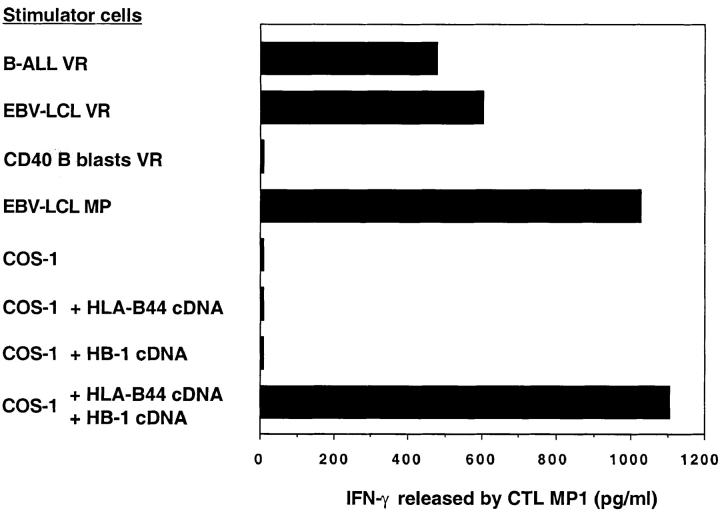
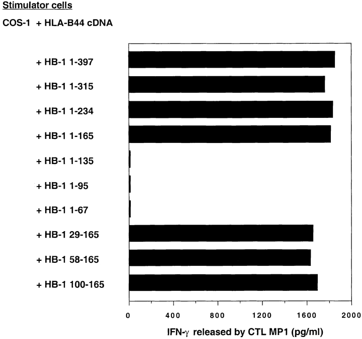
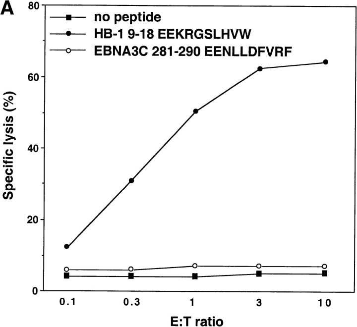
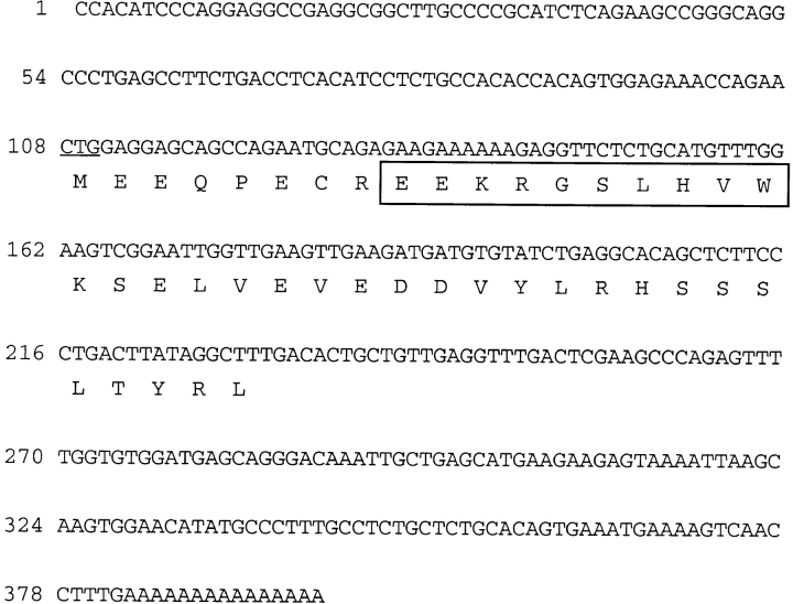
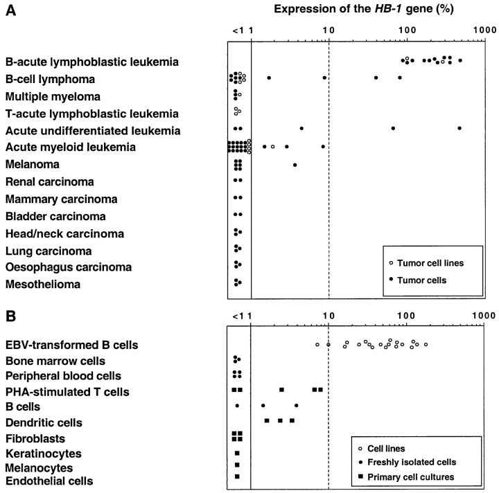
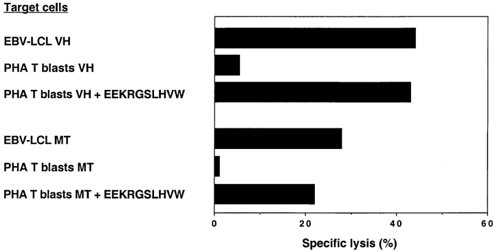
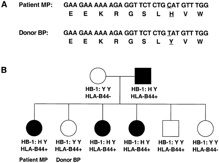
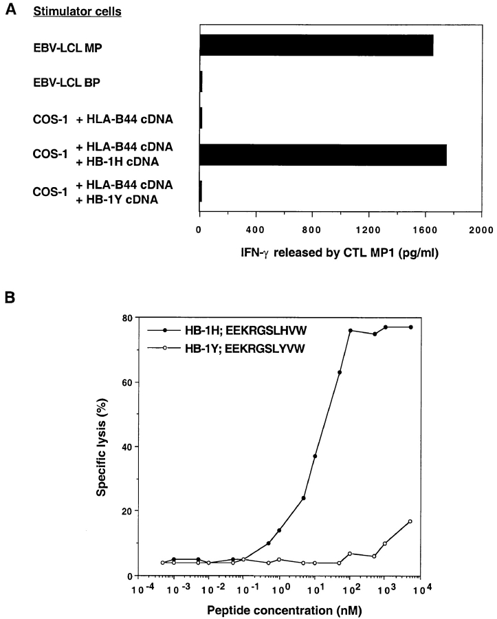
Human minor histocompatibility antigens (mHags) play an important role in the induction of cytotoxic T lymphocyte (CTL) reactivity against leukemia after human histocompatibility leukocyte antigen (HLA)-identical allogeneic bone marrow transplantation (BMT). As most mHags are not leukemia specific but are also expressed by normal tissues, antileukemia reactivity is often associated with life-threatening graft-versus-host disease (GVHD). Here, we describe a novel mHag, HB-1, that elicits donor-derived CTL reactivity in a B cell acute lymphoblastic leukemia (B-ALL) patient treated by HLA-matched BMT. We identified the gene encoding the antigenic peptide recognized by HB-1-specific CTLs. Interestingly, expression of the HB-1 gene was only observed in B-ALL cells and Epstein-Barr virus-transformed B cells. The HB-1 gene-encoded peptide EEKRGSLHVW is recognized by the CTL in association with HLA-B44. Further analysis reveals that a polymorphism in the HB-1 gene generates a single amino acid exchange from His to Tyr at position 8 within this peptide. This amino acid substitution is critical for recognition by HB-1-specific CTLs. The restricted expression of the polymorphic HB-1 Ag by B-ALL cells and the ability to generate HB-1-specific CTLs in vitro using peptide-loaded dendritic cells offer novel opportunities to specifically target the immune system against B-ALL without the risk of evoking GVHD.
Figures

Similar articles
-
Recognition of a B cell leukemia-associated minor histocompatibility antigen by CTL.J Immunol. 1997 Jan 15;158(2):560-5. J Immunol. 1997. PMID: 8992968
-
A single minor histocompatibility antigen encoded by UGT2B17 and presented by human leukocyte antigen-A*2902 and -B*4403.Transplantation. 2007 May 15;83(9):1242-8. doi: 10.1097/01.tp.0000259931.72622.d1. Transplantation. 2007. PMID: 17496542
-
Bi-directional allelic recognition of the human minor histocompatibility antigen HB-1 by cytotoxic T lymphocytes.Eur J Immunol. 2002 Oct;32(10):2748-58. doi: 10.1002/1521-4141(2002010)32:10<2748::AID-IMMU2748>3.0.CO;2-T. Eur J Immunol. 2002. PMID: 12355426
-
Alloreactivity and the predictive value of anti-recipient specific interleukin 2 producing helper T lymphocyte precursor frequencies for alloreactivity after bone marrow transplantation.Dan Med Bull. 2002 May;49(2):89-108. Dan Med Bull. 2002. PMID: 12064093 Review.
-
Human minor histocompatibility antigens: new concepts for marrow transplantation and adoptive immunotherapy.Immunol Rev. 1997 Jun;157:125-40. doi: 10.1111/j.1600-065x.1997.tb00978.x. Immunol Rev. 1997. PMID: 9255626 Review.
Cited by
-
NVX-412, a new oncology drug candidate, induces S-phase arrest and DNA damage in cancer cells in a p53-independent manner.PLoS One. 2012;7(9):e45015. doi: 10.1371/journal.pone.0045015. Epub 2012 Sep 13. PLoS One. 2012. PMID: 23028738 Free PMC article.
-
Identification of a polymorphic gene, BCL2A1, encoding two novel hematopoietic lineage-specific minor histocompatibility antigens.J Exp Med. 2003 Jun 2;197(11):1489-500. doi: 10.1084/jem.20021925. Epub 2003 May 27. J Exp Med. 2003. PMID: 12771180 Free PMC article.
-
Targeting alloreactive donor T-cells to hematopoietic system-restricted minor histocompatibility antigens to dissect graft-versus-leukemia effects from graft-versus-host disease after allogeneic stem cell transplantation.Int J Hematol. 2003 Oct;78(3):208-12. doi: 10.1007/BF02983796. Int J Hematol. 2003. PMID: 14604278 Review.
-
Bioinformatic analyses of mammalian 5'-UTR sequence properties of mRNAs predicts alternative translation initiation sites.BMC Bioinformatics. 2008 May 8;9:232. doi: 10.1186/1471-2105-9-232. BMC Bioinformatics. 2008. PMID: 18466625 Free PMC article.
-
Autosomal Minor Histocompatibility Antigens: How Genetic Variants Create Diversity in Immune Targets.Front Immunol. 2016 Mar 15;7:100. doi: 10.3389/fimmu.2016.00100. eCollection 2016. Front Immunol. 2016. PMID: 27014279 Free PMC article. Review.
References
-
- Horowitz MM, Gale RP, Sondel PM, Goldman JM, Kersey J, Kolb HJ, Rimm AA, Ringden O, Rozman C, Speck B, et al. Graft-versus-leukemia reactions after bone marrow transplantation. Blood. 1990;75:555–562. - PubMed
-
- Goulmy E. Human minor histocompatibility antigens. Curr Opin Immunol. 1996;8:75–81. - PubMed
-
- Perrault C, Roy DC, Fortin C. Immunodominant minor histocompatibility antigens: the major ones. Immunol Today. 1998;19:69–74. - PubMed
-
- Pardoll D. Taming the sinister side of BMT: Dr. Jekyll and Mr. Hyde. Nature Med. 1997;3:833–834. - PubMed
-
- Wallny HJ, Rammensee HG. Identification of classical minor histocompatibility antigen as cell-derived peptide. Nature. 1990;343:275–278. - PubMed
Publication types
MeSH terms
Substances
LinkOut - more resources
Full Text Sources
Other Literature Sources
Molecular Biology Databases
Research Materials
