. 1999 Jan 18;189(2):403-12.
doi: 10.1084/jem.189.2.403.

Lymphotoxin alpha/beta and tumor necrosis factor are required for stromal cell expression of homing chemokines in B and T cell areas of the spleen

Affiliations

Lymphotoxin alpha/beta and tumor necrosis factor are required for stromal cell expression of homing chemokines in B and T cell areas of the spleen

V N Ngo et al. J Exp Med. .

Abstract

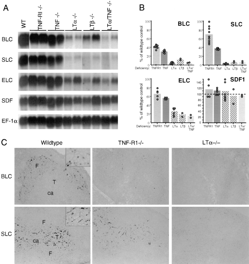
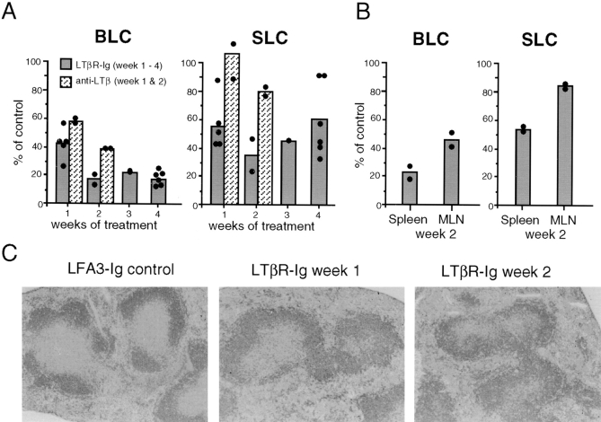
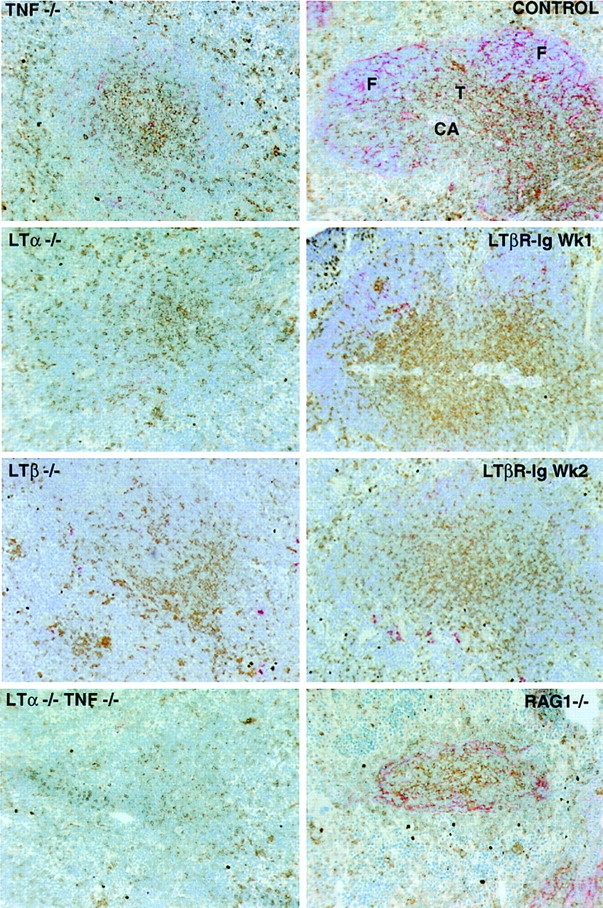
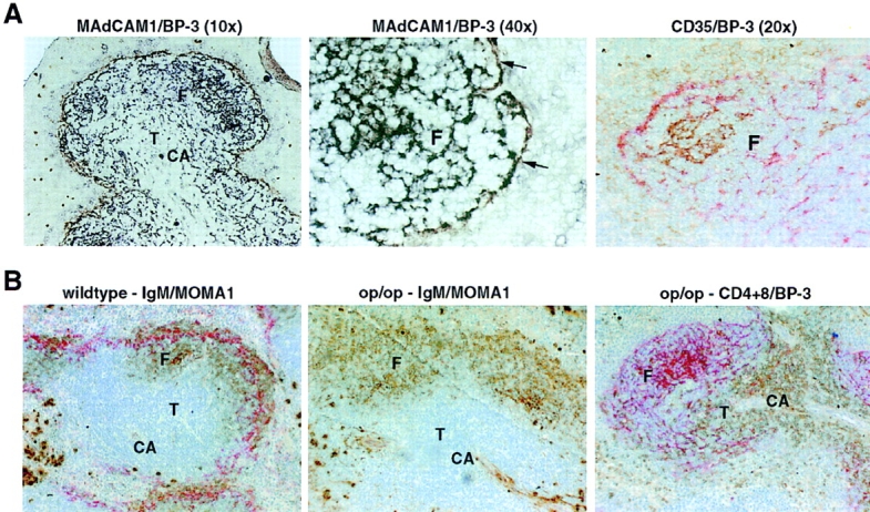
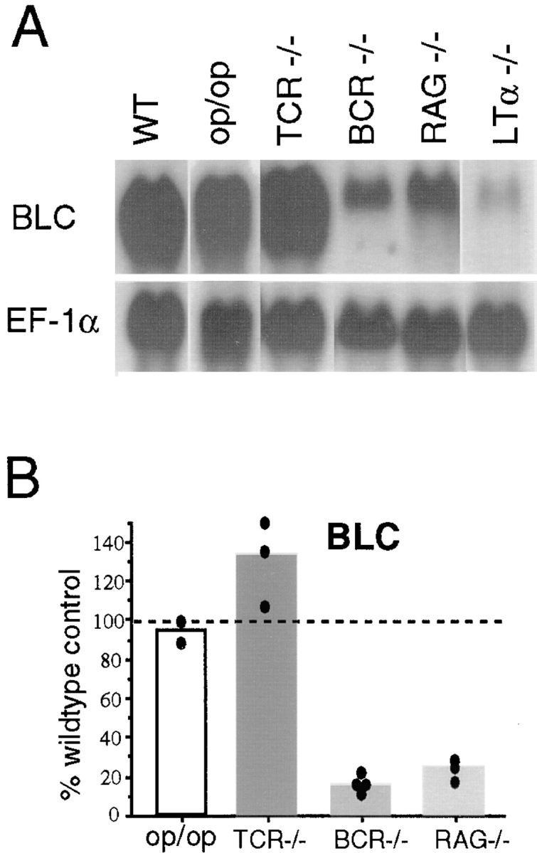
Mice deficient in the cytokines tumor necrosis factor (TNF) or lymphotoxin (LT) alpha/beta lack polarized B cell follicles in the spleen. Deficiency in CXC chemokine receptor 5 (CXCR5), a receptor for B lymphocyte chemoattractant (BLC), also causes loss of splenic follicles. Here we report that BLC expression by follicular stromal cells is defective in TNF-, TNF receptor 1 (TNFR1)-, LTalpha- and LTbeta-deficient mice. Treatment of adult mice with antagonists of LTalpha1beta2 also leads to decreased BLC expression. These findings indicate that LTalpha1beta2 and TNF have a role upstream of BLC/CXCR5 in the process of follicle formation. In addition to disrupted follicles, LT-deficient animals have disorganized T zones. Expression of the T cell attractant, secondary lymphoid tissue chemokine (SLC), by T zone stromal cells is found to be markedly depressed in LTalpha-, and LTbeta-deficient mice. Expression of the SLC-related chemokine, Epstein Barr virus-induced molecule 1 ligand chemokine (ELC), is also reduced. Exploring the basis for the reduced SLC expression led to identification of further disruptions in T zone stromal cells. Together these findings indicate that LTalpha1beta2 and TNF are required for the development and function of B and T zone stromal cells that make chemokines necessary for lymphocyte compartmentalization in the spleen.


Figures

Figure 1
Figure 1
Reduced expression of lymphoid tissue chemokines in TNF/TNFR1- and LTα/β-deficient mouse spleen. (A) Northern blot analysis of total RNA isolated from spleen tissue of the indicated mice and probed to detect expression of BLC, SLC, ELC, and SDF1. Hybridization to EF-1α was used to control for RNA loaded. For SDF1, the hybridization signals for SDF1α and SDF1β (reference 18) were similar and the signal for SDF1α is shown. WT, wild-type. (B) Relative chemokine mRNA levels as determined by PhosphorImager analysis of the Northern blot shown in A and additional blots, after correcting for differences in RNA loading from the corresponding EF-1α value. Data from individual mice are shown as open circles and means as shaded bars. (C) In situ hybridization analysis of BLC and SLC expression in spleen from wild-type, TNFR1-deficient, or LTα-deficient mice. Original magnification: ×10. ca, central arteriole; F, follicle; T, T zone. The insets in the BLC and SLC wild-type control panels are included to show the morphology of the chemokine-expressing stromal cells (original magnification: ×40).
Figure 2
Figure 2
Decreased BLC expression in mice treated with LTα1β2 antagonists. (A) Relative chemokine mRNA levels as determined by Northern blot and PhosphorImager analysis of total spleen RNA from mice treated for the indicated time period with LTβR-Ig (100 μg/wk, i.p.) or hamster anti-LTβ mAb (200 μg/wk, i.p.). Control mice for the LTβR-Ig treatment were treated with equal doses of LFA3-Ig, and controls for the mAb treatment were given hamster anti-KLH mAb. Each sample was corrected for differences in RNA loading using the value obtained with an EF-1α probe. Chemokine expression as percentage of control was calculated by dividing the corrected value for each treated mouse with the mean corrected value for the controls at that time point. Data from individual mice are shown as filled circles and means as shaded bars. (B) Relative chemokine mRNA levels in spleen and mesenteric lymph nodes from animals treated for 2 wk with LTβR-Ig (100 μg/wk, i.p.). Calculations were made as in A. (C) Disrupted follicular organization in LTβR-Ig–treated mice. Spleen tissue from mice treated with LFA3-Ig for 2 wk or LTβR-Ig for 1 or 2 wk was sectioned and stained with B220 (dark gray) to detect B cells.
Figure 3
Figure 3
Disruption of BP-3 expression in follicles and T zone of TNF-, TNFR1-, LTα-, and LTβ-deficient mice and LTβR-Ig–treated mice. Spleen tissue from the indicated mutant mice or mice treated with soluble LTβR-Ig for 1–2 wk or from a wild-type control was sectioned and stained to detect T cells (combination of anti-CD4 and anti-CD8; brown) and BP-3 (red). The CD4 and CD8 staining in the RAG-1−/− spleen does not represent T cells, as there was no staining for CD3 (not shown). CA, central arteriole; F, follicle; T, T zone. Original magnification: ×10.
Figure 4
Figure 4
Costaining of BP-3+ stromal cell subsets with MAdCAM-1 and CD35 (CR1) and normal follicular organization and BP-3 expression in op/op mice. (A) Spleen tissue from wild-type mice was sectioned and stained to detect MAdCAM-1 (brown) and BP-3 (black; left and center panels), or CD35 (brown) and BP-3 (red; right panel). Arrows in center panel indicate MAdCAM-1 and BP-3 double-stained cells. The faint brown CD35 staining corresponds to CD35high marginal zone B cells and CD35low follicular B cells. Original magnification: ×10, ×20, or ×40, as indicated. (B) Spleen tissue from wild-type (left) or op/op (center and right) mice was sectioned and stained to detect: IgM (brown) and MOMA1 (red; left and center), or CD4 and CD8 (brown) and BP-3 (red; right). Note the lack of MOMA1+ MMM staining in the op/op mutant. Original magnification: ×10. CA, central arteriole; F, follicle; T, T zone.
Figure 5
Figure 5
MZM independence and B lymphocyte dependence of BLC expression. (A) Northern blot analysis of total RNA isolated from spleen tissue of op/op, TCR-β−/−δ−/− (TCR−/−), μMT (BCR−/−), and RAG-1−/− mice, probed to detect expression of BLC and EF-1α. (B) Relative chemokine mRNA levels as determined by PhosphorImager analysis of the Northern blot shown in A and additional blots, after correcting for differences in RNA loading from the corresponding EF-1α value.

References

    1. Matsumoto M, Fu YX, Molina H, Chaplin DD. Lymphotoxin-alpha-deficient and TNF receptor-I- deficient mice define developmental and functional characteristics of germinal centers. Immunol Rev. 1997;156:137–144. - PubMed
    1. Futterer A, Mink K, Luz A, Kosco-Vilbois MH, Pfeffer K. The lymphotoxin β receptor controls organogenesis and affinity maturation in peripheral lymphoid tissues. Immunity. 1998;9:59–70. - PubMed
    1. Pasparakis M, Alexopoulou L, Episkopou V, Kollias G. Immune and inflammatory responses in TNF alpha–deficient mice: a critical requirement for TNF alpha in the formation of primary B cell follicles, follicular dendritic cell networks and germinal centers, and in the maturation of the humoral immune response. J Exp Med. 1996;184:1397–1411. - PMC - PubMed
    1. Neumann B, Machleidt T, Lifka A, Pfeffer K, Vestweber D, Mak TW, Holzmann B, Kronke M. Crucial role of 55-kilodalton TNF receptor in TNF-induced adhesion molecule expression and leukocyte organ infiltration. J Immunol. 1996;156:1587–1593. - PubMed
    1. Forster R, Mattis AE, Kremmer E, Wolf E, Brem G, Lipp M. A putative chemokine receptor, BLR1, directs B cell migration to defined lymphoid organs and specific anatomic compartments of the spleen. Cell. 1996;87:1–20. - PubMed

Publication types

MeSH terms