. 1999 Jan 18;189(2):423-34.
doi: 10.1084/jem.189.2.423.

Migration kinetics and final destination of type 1 and type 2 CD8 effector cells predict protection against pulmonary virus infection

Affiliations

Migration kinetics and final destination of type 1 and type 2 CD8 effector cells predict protection against pulmonary virus infection

A Cerwenka et al. J Exp Med. .

Abstract

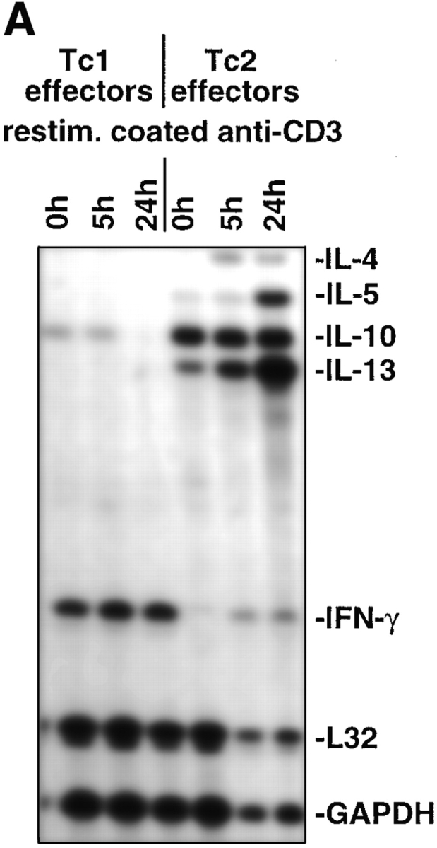
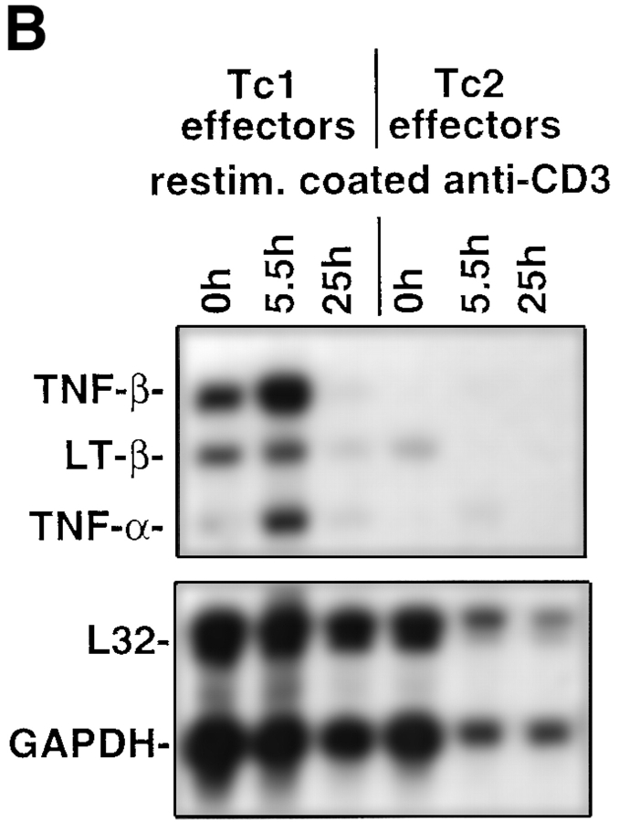
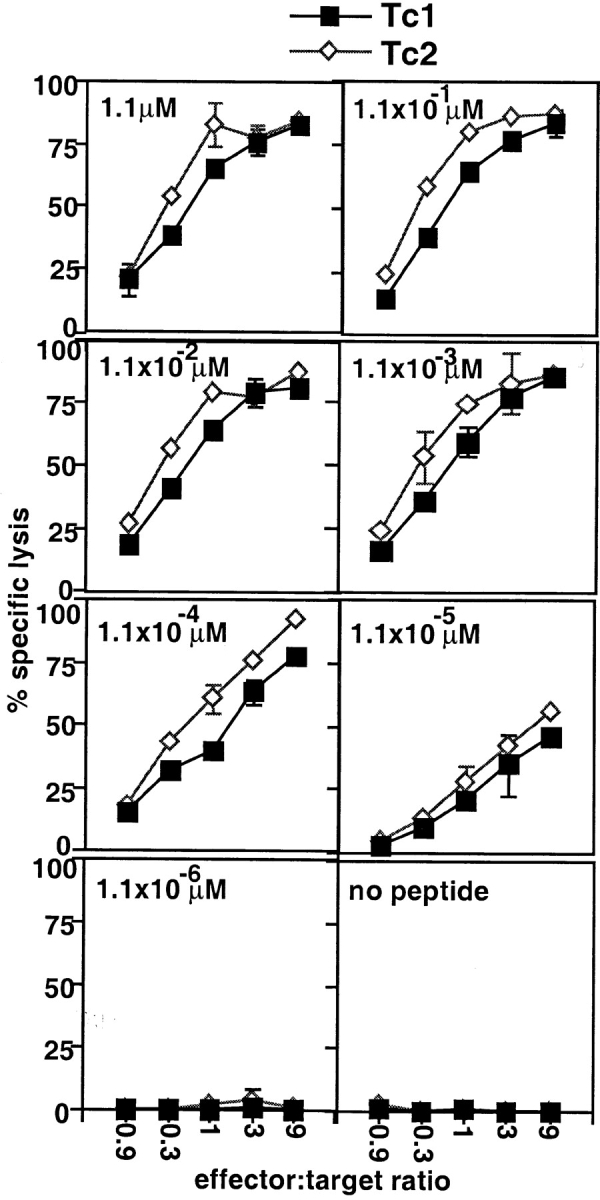
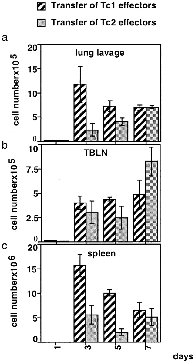
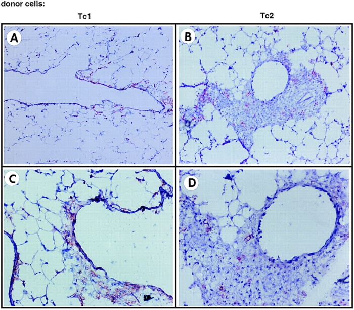
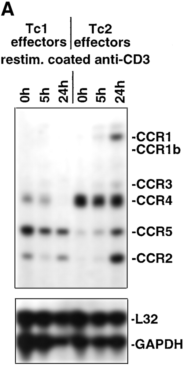
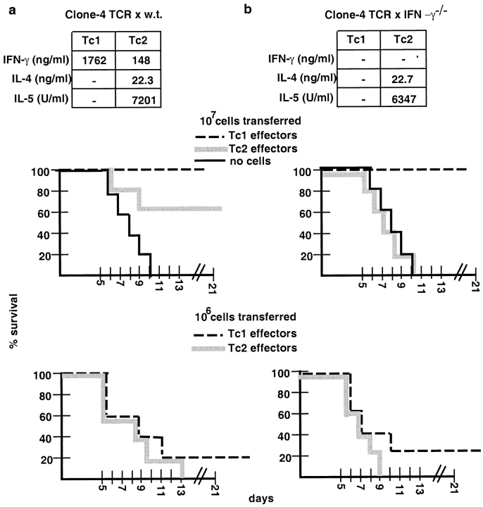
The requirements for CD8 T cells to provide protection against a localized virus infection in models of adoptive immunotherapy are not well defined. Here we investigated the protective value of defined in vitro-generated hemagglutinin (HA) peptide-specific primary CD8 T cell effectors from the clone 4 T cell receptor transgenic mice, secreting type 1 or type 2 cytokines, against pulmonary infection with whole influenza virus. Cytotoxic T lymphocytes producing type 1 and type 2 cytokine (Tc1 and Tc2) populations were equally cytolytic, but Tc1 effectors and not Tc2 effectors reduced the pulmonary virus titer early during infection. Host recovery mediated by Tc1 effectors was found to be independent of interferon gamma production. Tc2 effectors entered the lung with delayed kinetics as compared with Tc1 effectors, and after lung entry Tc2 effector cells did not localize near the infected airway epithelium as did Tc1 effectors but were found within clusters of inflammatory cells distant from the epithelium. We also show that the expression of several chemokine receptors was selectively regulated in the Tc1 and Tc2 subsets. Thus, the protective value of a CD8 cell population against pulmonary influenza virus infection is strongly correlated with its ability to exert its effector potential at the site of virus infection.


Figures

Figure 1
Figure 1
Cytokine mRNA expression within CD8 T cell subsets. On day 4 after in vitro culture RNA from 107 Tc1 and Tc2 effectors was prepared (t0) as described in Materials and Methods. Subsequently, 107 Tc1 or Tc2 effectors were restimulated with plate-bound anti-CD3 mAb (10 μg/ml) and RNA was prepared at the indicated time points. The RiboquantTM Multi-Probe protection assay mCK-1 (A) or mCK-3 (B) was used to detect cytokine mRNA. The results shown are representative for two independently performed experiments.
Figure 1
Figure 1
Cytokine mRNA expression within CD8 T cell subsets. On day 4 after in vitro culture RNA from 107 Tc1 and Tc2 effectors was prepared (t0) as described in Materials and Methods. Subsequently, 107 Tc1 or Tc2 effectors were restimulated with plate-bound anti-CD3 mAb (10 μg/ml) and RNA was prepared at the indicated time points. The RiboquantTM Multi-Probe protection assay mCK-1 (A) or mCK-3 (B) was used to detect cytokine mRNA. The results shown are representative for two independently performed experiments.
Figure 2
Figure 2
CTL activity of Tc1 and Tc2 effectors. The cytolytic activity of 4 d Tc1 (▪) and Tc2 effectors (⋄) was assayed in a 4-h 51Cr-release assay. P815 cells (104 cells/well) pulsed with titrated amounts of the HA peptide (1.1– 1.1 × 10⁻⁶ μM, indicated in the lefthand corners) were used as targets. The results are representative for three independently performed experiments.
Figure 3
Figure 3
Kinetics of migration of Tc1 and Tc2 effectors to the lung. 107 Tc1 and Tc2 effectors expressing Thy 1.2 were adoptively transferred into influenza infected Thy 1.1 BALB/c hosts and cells from lung lavages of recipients of Tc1 (left panels) or Tc2 (right panels) effectors were stained with Thy 1.2 PE and CD8 FITC in order to identify cells of donor origin on days 1, 3, 5, and 7. Dot-plots were gated on PI-negative live cells. The migration kinetics into the lung of one representative animal out of three is shown.
Figure 4
Figure 4
Absolute donor cell numbers recovered from lung lavage, spleen, and TBLN on days 1, 3, 5, and 7 after influenza virus infection and cell transfer. 107 Tc1 and Tc2 effectors expressing Thy 1.2 were adoptively transferred into influenza-infected Thy 1.1 BALB/c hosts as described in Fig. 3. To assess absolute donor cell numbers in the different organs, total cell counts from the isolated organs were performed and were multiplied by the percentages of donor cells determined by FACS® as shown in Fig. 3. Here, three animals per group were analyzed and the average donor cell numbers ± SE isolated from lung lavage (a), TBLN (b), and spleen (c) are shown. It should be noted that the adoptively transferred cells were injected at 4 PM on day 0 and the day 1 mice were killed at 7 AM, an interval of only 15 h.
Figure 5
Figure 5
Immunohistochemistry of lungs of recipients of Tc1 and Tc2 effector cells. 107 Tc1 (A and C) and Tc2 effectors (B and D) expressing Thy 1.2 were adoptively transferred into influenza-infected Thy 1.1 BALB/c hosts. On day 5 after infection and cell transfer, lungs were removed and frozen sections were prepared as described in Materials and Methods. Sections were stained with a biotinylated Thy 1.2 mAb or the respective biotinylated isotype control, and the color reaction was developed using a red substrate. The staining for Thy 1.2 (donor cells) was highly specific, because no red stain was detected when slides were incubated with the respective isotype control (data not shown). Original magnification: A and B, ×100; C and D, ×200. The results are representative of four independently performed experiments.
Figure 6
Figure 6
Chemokine receptor mRNA expression within CD8 T cell subsets. On day 4 after in vitro stimulation RNA from 107 Tc1 and Tc2 effectors was prepared (t0) as described in Materials and Methods before adoptive transfers. 107 Tc1 and Tc2 effectors were restimulated with plate-bound anti-CD3 mAb (10 μg/ml), and RNA was prepared at the indicated time points. The RiboQuantTM Multi-Probe protection assay mCR-5 (A) or mCR-6 (B) was used to detect chemokine receptor mRNA. The results shown are representative for three independently performed experiments.
Figure 6
Figure 6
Chemokine receptor mRNA expression within CD8 T cell subsets. On day 4 after in vitro stimulation RNA from 107 Tc1 and Tc2 effectors was prepared (t0) as described in Materials and Methods before adoptive transfers. 107 Tc1 and Tc2 effectors were restimulated with plate-bound anti-CD3 mAb (10 μg/ml), and RNA was prepared at the indicated time points. The RiboQuantTM Multi-Probe protection assay mCR-5 (A) or mCR-6 (B) was used to detect chemokine receptor mRNA. The results shown are representative for three independently performed experiments.
Figure 7
Figure 7
Phenotype of Tc1 and Tc2 donor cells upon re-isolation from infected hosts. 107 Tc1 and Tc2 effectors expressing Thy 1.2 were adoptively transferred into influenza-infected Thy 1.1 BALB/c hosts. On day 5 after infection and cell transfer, TBLN (a) or lung lavages (b) of recipients of Tc1 or Tc2 effectors were double stained with FITC-conjugated mAbs directed against CD44, Ly6C, and CD62L, respectively and with PE-conjugated Thy 1.2 mAb analyzed on a FACScan®. Histograms were gated on Thy 1.2–positive and PI-negative live cells. Data are representative of three similar experiments performed.
Figure 8
Figure 8
(a) Tc1 and Tc2 effectors retain their polarization profile during influenza infection in vivo. Tc1 and Tc2 effectors (107) were adoptively transferred into influenza-infected hosts. On day 4 after infection and cell transfer, CD8 T cells were enriched from the lung digest and similar numbers of Tc1 and Tc2 donor cells (Thy 1.2–positive cells) were restimulated with P815 loaded with the HA peptide (11 μM). Supernatants were collected after 24 h and cytokines were measured by specific ELISAs. No cytokines were detected when Tc1 and Tc2 effectors were restimulated in the absence of the HA peptide (data not shown). No cytokines were detected when the cell cultures were depleted of Thy 1.2 (donor cells)–positive cells, indicating that the observed cytokine production was due to the adoptively transferred Thy 1.2 donor cell population (data not shown). (b) CTL activity of Tc1 and Tc2 effectors upon reisolation from infected lung tissue. 107 Tc1 (▪) and Tc2 effectors (□) were adoptively transferred into influenza-infected hosts. On day 4 after infection and cell transfer, CD8 T cells were enriched from the lung digest and similar numbers of Tc1 and Tc2 donor cells (Thy 1.2– positive cells) were assayed in a 4-h 51Cr-release assay using P815 cells (104 cells/well) pulsed with the HA peptide (11 μM) as targets. Only little CTL activity was detected, and the effectors were depleted of Thy 1.2 (donor cells)–positive cells (♦) in vitro before the CTL assay, indicating that the observed CTL activity was indeed due to the adoptively transferred Thy 1.2 donor cell population.
Figure 8
Figure 8
(a) Tc1 and Tc2 effectors retain their polarization profile during influenza infection in vivo. Tc1 and Tc2 effectors (107) were adoptively transferred into influenza-infected hosts. On day 4 after infection and cell transfer, CD8 T cells were enriched from the lung digest and similar numbers of Tc1 and Tc2 donor cells (Thy 1.2–positive cells) were restimulated with P815 loaded with the HA peptide (11 μM). Supernatants were collected after 24 h and cytokines were measured by specific ELISAs. No cytokines were detected when Tc1 and Tc2 effectors were restimulated in the absence of the HA peptide (data not shown). No cytokines were detected when the cell cultures were depleted of Thy 1.2 (donor cells)–positive cells, indicating that the observed cytokine production was due to the adoptively transferred Thy 1.2 donor cell population (data not shown). (b) CTL activity of Tc1 and Tc2 effectors upon reisolation from infected lung tissue. 107 Tc1 (▪) and Tc2 effectors (□) were adoptively transferred into influenza-infected hosts. On day 4 after infection and cell transfer, CD8 T cells were enriched from the lung digest and similar numbers of Tc1 and Tc2 donor cells (Thy 1.2– positive cells) were assayed in a 4-h 51Cr-release assay using P815 cells (104 cells/well) pulsed with the HA peptide (11 μM) as targets. Only little CTL activity was detected, and the effectors were depleted of Thy 1.2 (donor cells)–positive cells (♦) in vitro before the CTL assay, indicating that the observed CTL activity was indeed due to the adoptively transferred Thy 1.2 donor cell population.
Figure 9
Figure 9
Recipient survival after adoptive transfer of Tc1 and Tc2 effector cells into lethally influenza-infected hosts. Tc1 (dashed line) and Tc2 effectors (gray line) generated from the clone 4 TCR transgenic mice on the wild-type (a) or on the IFN-γ−/− background (b) were prepared as described in Materials and Methods. Cytokine production of the effector populations before adoptive transfer is indicated. On day 4 of primary stimulation, cells were washed and 107 (center panel) or 106 (bottom panel) effector cells were adoptively transferred into sex- and age-matched BALB/c recipients, which were infected intranasally with 10 LD50 of the influenza virus PR8, approximately 1 h before the cell transfer. As a control no cells were injected into influenza-infected hosts (thin line) and all recipient animals died. The percentage of survival of five animals per group is presented for the time points indicated. Survival was monitored for 21 days. Data presented are representative for 4 (a) or 2 (b) independently performed experiments.

References

    1. Doherty PC, Topham DJ, Tripp RA, Cardin RD, Brooks JW, Stevenson PG. Effector CD4+ and CD8+ T-cell mechanisms in the control of respiratory virus infections. Immunol Rev. 1997;159:105–117. - PubMed
    1. Gerhard W, Mozdzanowska K, Furchner M, Washko G, Maiese K. Role of the B-cell response in recovery of mice from primary influenza virus infection. Immunol Rev. 1997;159:95–103. - PubMed
    1. Yap KL, Ada GL, McKenzie IFC. Transfer of specific cytotoxic lymphocytes protects mice inoculated with influenza virus. Nature. 1978;238:238–239. - PubMed
    1. Lukacher AE, Braciale VL, Braciale TJ. In vivo function of influenza virus-specific cytotoxic T lymphocyte clones is highly specific. J Exp Med. 1984;160:814–826. - PMC - PubMed
    1. Topham DJ, Tripp RA, Doherty PC. CD8+ T cells clear influenza virus by perforin or Fas-dependent processes. J Immunol. 1997;159:5197–5200. - PubMed

Publication types

MeSH terms