. 1999 Jan 19;96(2):371-5.
doi: 10.1073/pnas.96.2.371.

Antitumor activity of mannan-binding protein in vivo as revealed by a virus expression system: mannan-binding proteindependent cell-mediated cytotoxicity

Affiliations

Antitumor activity of mannan-binding protein in vivo as revealed by a virus expression system: mannan-binding proteindependent cell-mediated cytotoxicity

Y Ma et al. Proc Natl Acad Sci U S A. .

Abstract

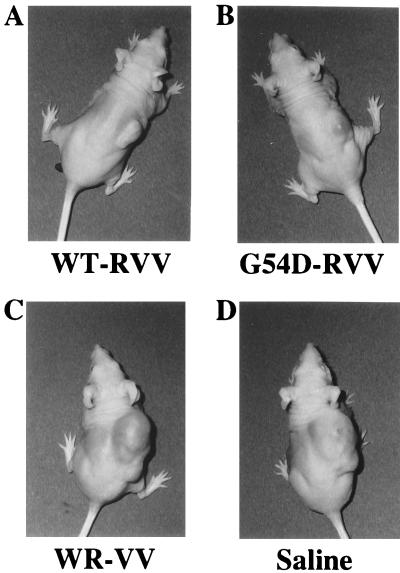
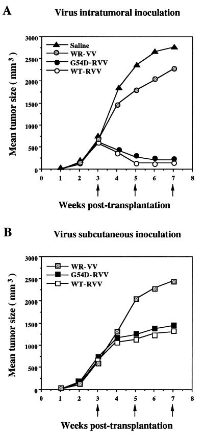
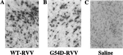
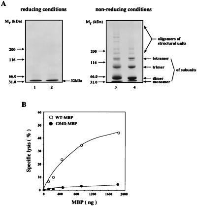
Mannan-binding protein (MBP), a Ca2+-dependent mammalian lectin specific for mannose and N-acetylglucosamine, is an important serum component associated with innate immunity. MBP activates complement and functions as a direct opsonin on binding to mannooligosaccharide-bearing pathogens. We have found that MBP recognizes and binds specifically to oligosaccharide ligands expressed on the surfaces of a human colorectal carcinoma. Interestingly, the recombinant vaccinia virus carrying human MBP gene was demonstrated to possess a potent growth-inhibiting activity against human colorectal carcinoma cells transplanted in KSN nude mice when administered by intratumoral or subcutaneous injection. Moreover, a significant prolongation of life span of tumor-bearing mice resulted from the treatment. This effect appears to be a consequence of local production of MBP. Unexpectedly, the mutant MBP, which had essentially no complement-activating activity, was nearly as active as wild-type MBP. These results indicated that MBP has a previously undescribed cytotoxic activity, which we propose to term MBP-dependent cell-mediated cytotoxicity(MDCC). In addition, this study provides a model for the development of an effective and specific host defense factor for cancer gene therapy.


Figures

Figure 1
Figure 1
Expression of the MBP ligands on human colorectal carcinoma SW1116 cell surfaces. The cultured SW1116 cells were incubated with human MBPs, followed by immunofluorescent staining with anti-human MBP mAb YM304 and with FITC-conjugated anti-mouse IgG. The cells were incubated with either WT-MBP (A, CF) or G54D-MBP (B) in the presence of 10 mM Ca2+ (A and B), 10 mM EDTA (C), 20 mM mannose/10 mM Ca2+ (D), 20 mM GalNAc/10 mM Ca2+ (E), and 20 mM GlcNAc/10 mM Ca2+ (F).
Figure 2
Figure 2
Photographs of SW1116 tumor-bearing KSN nude mice treated with the recombinant vaccinia virus carrying MBP genes. The pictures were taken 48 days after tumor transplantation. The tumor-bearing mice were inoculated intratumorally with two consecutive doses of WT-RVV (A), G54D-RVV (B), WR-VV control (C), or saline control (D).
Figure 3
Figure 3
Time course of SW1116 tumor growth inhibition by the recombinant vaccinia virus carrying MBP genes. Four different groups of SW1116 tumor-bearing KSN nude mice were inoculated intratumorally (A) or subcutaneously (B) with three consecutive doses of 5 × 10⁶ pfu of WT-RVV, G54D-RVV, WR-VV control, or saline control (arrows). Tumor size was measured in three dimensions every 3–4 days with Vernier calipers. Differences in tumor size were statistically analyzed with the Kruskal–Wallis test and the Wilcoxon test.
Figure 4
Figure 4
Immunohistochemistry of SW1116 tumors from tumor-bearing mice treated with the recombinant vaccinia virus carrying MBP genes. Tumor specimens inoculated intratumorally with three consecutive doses of the recombinant virus were stained with HRP-conjugated anti-human MBP mAb YM304. WT-RVV (A), G54D-RVV (B), and saline control (C).
Figure 5
Figure 5
(A) SDS/PAGE of WT- and G54D-MBPs expressed in SW1116 cells. Samples were electrophoresed on a 3–10% polyacrylamide gel according to the method of Laemmli (21) under reducing (Left) and nonreducing (Right) conditions. Proteins were stained with Coomassie Brilliant Blue R250. The positions of the molecular markers (kDa) are indicated on the left, and the arrows on the right indicate the positions of MBP monomers or oligomers of the subunit and the structural units. WT-MBP, lanes 1 and 3; G54D-MBP, lanes 2 and 4. (B) Dose dependence of the complement activation assay of wild-type and mutant G54D-MBPs expressed in SW1116 cells by passive hemolysis. Mannan-coated sheep erythrocytes were sensitized with 0–2,000 ng of recombinant wild-type or mutant G54D-MBPs and lysed with the guinea pig complement (2CH50).

References

    1. Kawasaki N, Kawasaki T, Yamashina I. J Biochem (Tokyo) 1983;94:937–947. - PubMed
    1. Kurata H, Sannoh T, Kozutsumi Y, Yokota Y, Kawasaki T. J Biochem (Tokyo) 1994;115:1148–1154. - PubMed
    1. Drickamer K, Dordal M S, Reynolds L. J Biol Chem. 1986;261:6878–6887. - PubMed
    1. Thiel S, Vorup-Jensen T, Stover C M, Schwaeble W, Laursen S B, Poulsen K, Willis A C, Eggleton P, Hansen S, Holmskov U, et al. Nature (London) 1997;386:506–510. - PubMed
    1. Childs R A, Drickamer K, Kawasaki T, Thiel S, Mizuochi T, Feizi T. Biochem J. 1989;262:131–138. - PMC - PubMed