

Myosin I heavy chain kinase: cloning of the full-length gene and acidic lipid-dependent activation by Rac and Cdc42
- PMID: 9892644
- PMCID: PMC15147
- DOI: 10.1073/pnas.96.2.394
Myosin I heavy chain kinase: cloning of the full-length gene and acidic lipid-dependent activation by Rac and Cdc42
Abstract
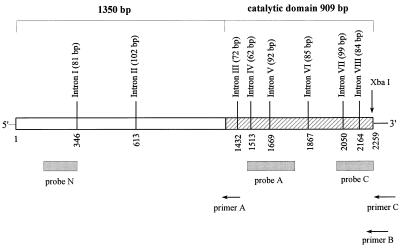
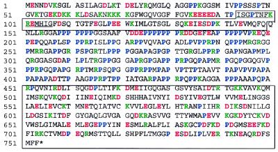
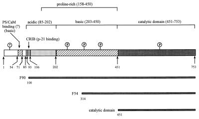
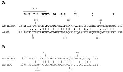
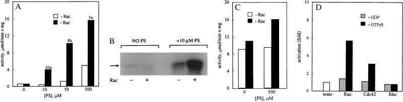
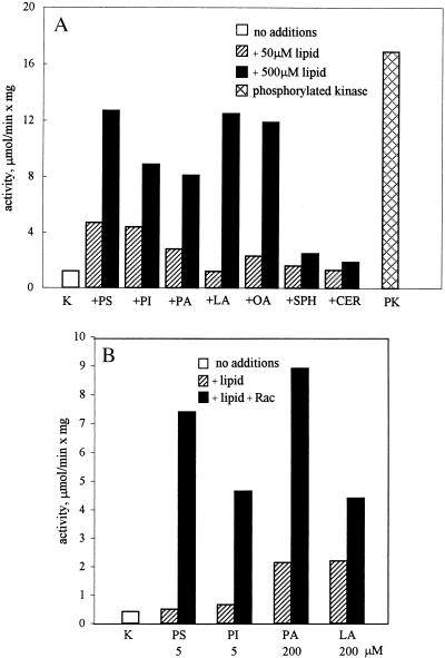
Acanthamoeba myosin I heavy chain kinase (MIHCK) phosphorylates the heavy chains of amoeba myosins I, increasing their actin-activated ATPase activities. The activity of MIHCK is increased by binding to acidic phospholipids or membranes and by autophosphorylation at multiple sites. Phosphorylation at a single site is necessary and sufficient for full activation of the expressed catalytic domain. The rate of autophosphorylation of native MIHCK is controlled by a region N-terminal to the catalytic domain. By its substrate specificity and the sequence of its C-terminal catalytic domain, MIHCK was identified as a p21-activated kinase (PAK). We have now cloned the full-length genomic DNA and cDNA of MIHCK and have shown it to contain the conserved p21-binding site common to many members of the PAK family. Like some mammalian PAKs, MIHCK is activated by Rac and Cdc42, and this activation is GTP-dependent and accompanied by autophosphorylation. In contrast to mammalian PAKs, activation of MIHCK by Rac and Cdc42 requires the presence of acidic lipids. Also unlike mammalian PAK, MIHCK is not activated by sphingosine or other non-negatively charged lipids. The acidic lipid-binding site is near the N terminus followed by the p21-binding region. The N-terminal regulatory domain of MIHCK contains alternating strongly positive and strongly negative regions. and the extremely Pro-rich middle region of MIHCK has a strongly acidic N-terminal segment and a strongly basic C-terminal segment. We propose that autophosphorylation activates MIHCK by neutralizing the basic segment of the Pro-rich region, thus unfolding the regulatory domain and abolishing its inhibition of the catalytic domain.
Figures
Similar articles
-
Calmodulin-binding and autoinhibitory domains of Acanthamoeba myosin I heavy chain kinase, a p21-activated kinase (PAK).J Biol Chem. 2001 Dec 14;276(50):47468-73. doi: 10.1074/jbc.M108957200. Epub 2001 Sep 28. J Biol Chem. 2001. PMID: 11579107
-
Cloning and characterization of a Dictyostelium myosin I heavy chain kinase activated by Cdc42 and Rac.J Biol Chem. 1996 Oct 25;271(43):27044-8. doi: 10.1074/jbc.271.43.27044. J Biol Chem. 1996. PMID: 8900194
-
p21-activated kinase has substrate specificity similar to Acanthamoeba myosin I heavy chain kinase and activates Acanthamoeba myosin I.Proc Natl Acad Sci U S A. 1997 Feb 18;94(4):1092-5. doi: 10.1073/pnas.94.4.1092. Proc Natl Acad Sci U S A. 1997. PMID: 9037011 Free PMC article.
-
Regulation of Dictyostelium myosin I and II.Biochim Biophys Acta. 2001 Mar 15;1525(3):245-61. doi: 10.1016/s0304-4165(01)00110-6. Biochim Biophys Acta. 2001. PMID: 11257438 Review.
-
The p21Rac/Cdc42-activated kinases (PAKs).Int J Biochem Cell Biol. 1998 Aug;30(8):857-62. doi: 10.1016/s1357-2725(98)00059-4. Int J Biochem Cell Biol. 1998. PMID: 9744077 Review.
Cited by
-
Regulation of nonmuscle myosins by heavy chain phosphorylation.J Muscle Res Cell Motil. 2001;22(2):163-73. doi: 10.1023/a:1010552929028. J Muscle Res Cell Motil. 2001. PMID: 11519739 Review.
-
Direct involvement of yeast type I myosins in Cdc42-dependent actin polymerization.J Cell Biol. 2000 Jan 24;148(2):363-73. doi: 10.1083/jcb.148.2.363. J Cell Biol. 2000. PMID: 10648569 Free PMC article.
-
PAK family kinases: Physiological roles and regulation.Cell Logist. 2012 Apr 1;2(2):59-68. doi: 10.4161/cl.21912. Cell Logist. 2012. PMID: 23162738 Free PMC article.
-
Cdc42/Rac Interactive Binding Containing Effector Proteins in Unicellular Protozoans With Reference to Human Host: Locks of the Rho Signaling.Front Genet. 2022 Feb 2;13:781885. doi: 10.3389/fgene.2022.781885. eCollection 2022. Front Genet. 2022. PMID: 35186026 Free PMC article. Review.
-
An experimentally based computer search identifies unstructured membrane-binding sites in proteins: application to class I myosins, PAKS, and CARMIL.J Biol Chem. 2010 Feb 19;285(8):5738-47. doi: 10.1074/jbc.M109.066910. Epub 2009 Dec 15. J Biol Chem. 2010. PMID: 20018884 Free PMC article.
References
-
- Pollard T D, Korn E D. J Biol Chem. 1973;248:4691–4697. - PubMed
-
- Maruta H, Korn E D. J Biol Chem. 1977;252:8329–8332. - PubMed
-
- Brzeska H, Lynch T J, Martin B M, Korn E D. J Biol Chem. 1989;264:19340–19348. - PubMed
-
- Lynch T J, Brzeska H, Miyata H, Korn E D. J Biol Chem. 1989;264:19333–19339. - PubMed
-
- Brzeska H, Korn E D. J Biol Chem. 1996;271:16983–16986. - PubMed
MeSH terms
Substances
Associated data
- Actions
- Actions
LinkOut - more resources
Full Text Sources
Miscellaneous
