. 1999 Jan 19;96(2):429-34.
doi: 10.1073/pnas.96.2.429.

Involvement of regulatory and catalytic subunits of phosphoinositide 3-kinase in NF-kappaB activation

Affiliations

Involvement of regulatory and catalytic subunits of phosphoinositide 3-kinase in NF-kappaB activation

C Béraud et al. Proc Natl Acad Sci U S A. .

Abstract

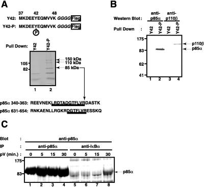
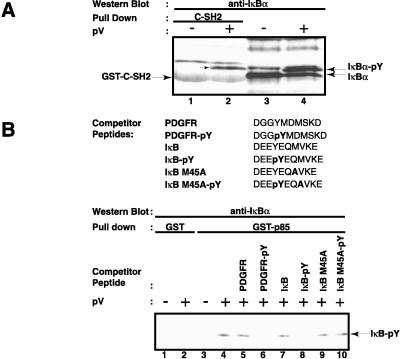
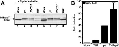
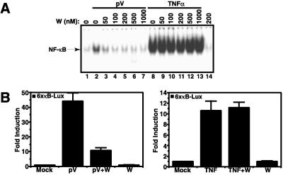
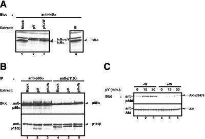
Hypoxia, reoxygenation, and the tyrosine phosphatase inhibitor pervanadate activate the transcription factor NF-kappaB, involving phosphorylation of its inhibitor IkappaB-alpha on tyrosine 42. This modification does not lead to degradation of IkappaB by the proteasome/ubiquitin pathway, as is seen on stimulation of cells with proinflammatory cytokines. It is currently unknown how tyrosine-phosphorylated IkappaB is removed from NF-kappaB. Here we show that p85alpha, the regulatory subunit of PI3-kinase, specifically associates through its Src homology 2 domains with tyrosine-phosphorylated IkappaB-alpha in vitro and in vivo after stimulation of T cells with pervanadate. This association could provide a mechanism by which newly tyrosine-phosphorylated IkappaB is sequestered from NF-kappaB. Another mechanism by which PI3-kinase contributed to NF-kappaB activation in response to pervanadate appeared to involve its catalytic p110 subunit. This was evident from the inhibition of pervanadate-induced NF-kappaB activation and reporter gene induction by treatment of cells with nanomolar amounts of the PI3-kinase inhibitor wortmannin. The compound had virtually no effect on tumor necrosis factor- and interleukin-1-induced NF-kappaB activities. Wortmannin did not inhibit tyrosine phosphorylation of IkappaB-alpha or alter the stability of the PI3-kinase complex but inhibited Akt kinase activation in response to pervanadate. Our data suggest that both the regulatory and the catalytic subunit of PI3-kinase play a role in NF-kappaB activation by the tyrosine phosphorylation-dependent pathway.


Figures

Figure 1
Figure 1
Proteins interacting with a tyrosine-phosphorylated IκB-α peptide. (A) Affinity purification. The sequence of the peptides used for the affinity purification from 293 cell extracts is shown on top. Amino acid residues 37–48 of IκB-α were fused to four glycine residues and a Flag epitope at the C terminus. Tyrosine 42 was either phosphorylated (Y42-P) or not (Y42). Proteins interacting with the nonphosphorylated peptide (lane 1) or the phosphorylated peptide (lane 2) were analyzed by SDS polyacrylamide gel electrophoresis and Coomassie blue staining. The position of two molecular mass markers is indicated on the left; the position of three proteins interacting only with the tyrosine-phosphorylated peptide is indicated on the right. Protein sequences of two tryptic peptides obtained from the 85-kDa band are shown underlined. They are found within the indicated regions of human PI3-kinase regulatory p85α subunit. (B) Both PI3-kinase subunits bind to the IκB-α peptide. Proteins from Jurkat T cells binding to the nonphosphorylated peptide (lanes 1 and 3) or tyrosine-phosphorylated IκB-α peptide (lanes 2 and 4) were analyzed by Western blotting using an antibody directed against the regulatory p85α subunit of PI3-kinase (lanes 1 and 2) or the catalytic p110β subunit of PI3-kinase (lanes 3 and 4). The positions of p85α and p110β are indicated by arrows on the right; the positions of four molecular mass markers on the left. (C) Interaction of endogenous p85α with endogenous IκB-α on pV treatment. Jurkat T cells (1.5 × 10⁶) pretreated with 400 nM wortmannin were induced with 400 μM pV for 0 (lanes 1 and 5), 5 (lanes 2 and 6), 15 (lanes 3 and 7), and 30 min (lanes 4 and 8). Cytosolic extracts were prepared and immunoprecipitated with anti-p85α (lanes 1–4) or anti-IκB-α (lanes 5–8). The presence of p85α in the complexes was detected by Western blotting. The position of p85α is indicated by an arrow.
Figure 2
Figure 2
Specificity of the interaction of tyrosine-phosphorylated IκB-α with the regulatory p85α subunit of PI3-kinase. (A) Interaction of tyrosine-phosphorylated IκB-α with the C-terminal SH2 domain of p85α. Two μg of GST-C-SH2 agarose conjugate (lanes 1 and 2) were incubated with whole-cell extracts from control (−) or 200 μM pV-treated (+) Jurkat T cells. The binding of IκB-α to the GST proteins was detected by Western blotting. The analysis of total cell extract by Western blotting for IκB-α is shown in lanes 3 and 4. The positions of IκB-α and its tyrosine-phosphorylated form, as well as that of the GST fusion protein, are indicated by arrows. (B) Specificity of interaction of tyrosine-phosphorylated IκB-α with p85α. Whole-cell extracts from control (−) and pV-treated (+) Jurkat T cells were incubated with 6 μM of the indicated peptides. Two μg of GST (lanes 1 and 2) or GST-p85α (lanes 3–10) agarose conjugates were added and the presence of IκB-α in the complexes was analyzed by Western blotting. The position of tyrosine-phosphorylated IκB-α is indicated.
Figure 3
Figure 3
The effect of TNFα on pV-induced events. (A) Protection of tyrosine-phosphorylated IκB-α from degradation through the TNFα pathway. Jurkat T cells were pretreated (lanes 1–4) or not (lanes 5–8) with 10 μg⋅ml−1 cycloheximide for 30 min, treated with 1 mM pV (lanes 2, 3, 6 and 7) and 10 min later induced with 50 ng⋅ml−1 TNFα (lanes 3, 4, 7 and 8) for 1 h. Whole-cell extracts were prepared and IκB-α was detected by Western blotting. The positions of IκB-α and its tyrosine-phosphorylated form are indicated. (B) pV does not impair TNFα signaling. Jurkat cells were transiently transfected with a plasmid containing a luciferase reporter gene driven by three repeats of the HIV type 1 (HIV-1) κB enhancer. Forty-eight hours after transfection, cells were treated with 1 mM pV and 1 h later induced with 50 ng⋅ml−1 TNFα for 5 h. Luciferase activities were measured and normalized on the basis of β-galactosidase expression from cotransfected pRSV-β-galactosidase. The values shown are averages (mean + SEM) of one representative experiment in which each transfection was performed in duplicate.
Figure 4
Figure 4
PI3-kinase activity is necessary for NF-κB induction by pV. (A) Effect of wortmannin on NF-κB DNA-binding activity. Jurkat cells were pretreated for 1 h with the indicated concentrations of wortmannin (W) and were stimulated with 1 mM pV (lanes 2–7) or 50 ng⋅ml−1 TNFα (lanes 8–13) for 1 h. Nuclear extracts were prepared and electrophoretic mobility shift assays were performed by using the IL-2 gene κB site as probe. The position of the NF-κB complex is indicated by an arrow. (B) Effect of wortmannin on the activation of a NF-κB-dependent reporter gene activity. Jurkat T cells were transiently transfected with a plasmid containing the luciferase reporter gene driven by three repeats of the HIV-1 κB enhancer. Forty-eight hours after transfection, cells were pretreated with 200 nM wortmannin for 1 h and then induced with 1 mM pV or 50 ng⋅ml−1 TNFα for 6 h, as indicated. Luciferase activities were measured and normalized to the protein concentration in each extract. The values shown are averages (mean + SEM) of one representative experiment in which each transfection was performed in quadruplicate.
Figure 5
Figure 5
Wortmannin does not perturb tyrosine phosphorylation of IκB-α and PI3-kinase heterodimer formation. (A) Effect of wortmannin on pV-mediated tyrosine phosphorylation of IκB-α. Jurkat T cells were pretreated with 200 nM of wortmannin (lanes 3 and 4) and induced with 200 μM pV (lanes 2 and 3) for 1 h. IκB-α phosphorylation status was analyzed by Western blotting. The position of IκB-α and tyrosine phosphorylated IκB-α is indicated. (B) Effect of wortmannin on PI3-kinase heterodimer formation. Jurkat T cells were pretreated with 200 nM of wortmannin (lanes 3 and 6) and were subsequently induced with 1 mM pV (lanes 2, 3, 5, 6) for 1 h. Cytosolic extracts were prepared and immunoprecipitated with anti-p85α (lanes 1–3) or anti-p110β (lanes 4–6). The presence of p85α (Upper) and p110β (Lower) in the immunocomplexes was analyzed by Western blotting. The positions of p85α and p110β are indicated by arrows. (C) pV induces PI3-kinase activity as measured by Akt serine 473 phosphorylation. Jurkat cells were pretreated (lanes 4–6) or not (lanes 1–3) with 100 nM of wortmannin and induced with 400 μM pV for 0 (lanes 1 and 4), 15 (lanes 2 and 5), or 30 min (lanes 3 and 6). The phosphorylation status of Akt was analyzed by Western blotting with an antibody specific for phosphorylated Akt (Upper). Total levels of Akt were measured with an anti-Akt antibody (Lower). The positions of phosphoserine 473-Akt and Akt are indicated by arrows.

References

    1. Baeuerle P A, Baltimore D. Cell. 1996;87:13–20. - PubMed
    1. Baldwin A S., Jr Annu Rev Immunol. 1996;14:649–683. - PubMed
    1. Verma I M, Stevenson J K, Schwarz E M, Van Antwerp D, Miyamoto S. Genes Dev. 1995;9:2723–2735. - PubMed
    1. Bours V, Villalobos J, Burd P R, Kelly K, Siebenlist U. Nature (London) 1990;348:76–80. - PubMed
    1. Ghosh S, Gifford A M, Riviere L R, Tempst P, Nolan G P, Baltimore D. Cell. 1990;62:1019–1029. - PubMed

LinkOut - more resources