. 1999 Jan 19;96(2):517-22.
doi: 10.1073/pnas.96.2.517.

Moe1, a conserved protein in Schizosaccharomyces pombe, interacts with a Ras effector, Scd1, to affect proper spindle formation

Affiliations

Moe1, a conserved protein in Schizosaccharomyces pombe, interacts with a Ras effector, Scd1, to affect proper spindle formation

C R Chen et al. Proc Natl Acad Sci U S A. .

Abstract

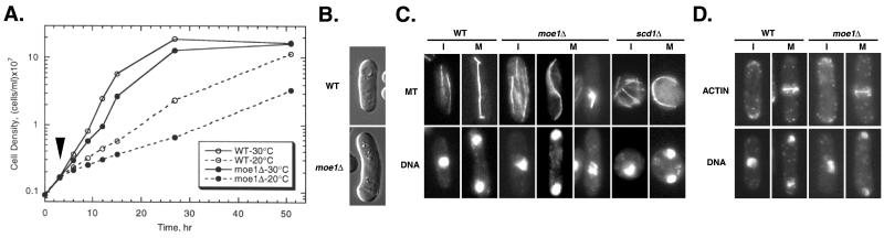
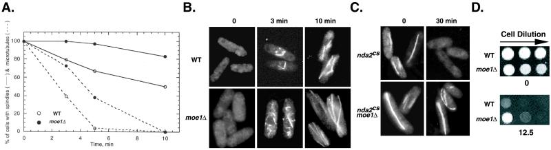
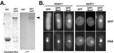
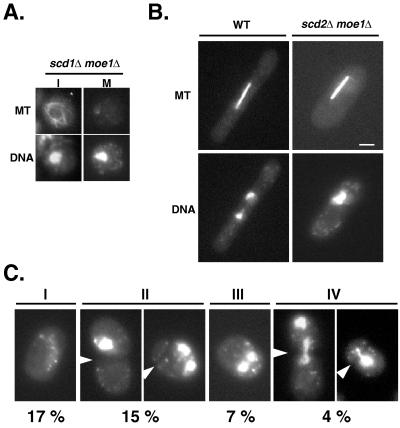
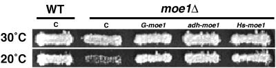
In fission yeast, Scd1/Ral1 is a putative guanine nucleotide exchange factor for Cdc42sp and also acts as a Ras1 effector necessary for the regulation of cytoskeleton organization. In this study, we have characterized a protein, Moe1, that binds directly to Scd1. A moe1 null (Delta) mutant exhibits numerous phenotypes indicative of abnormal microtubule functioning, including an abnormality in the spindle. moe1Delta mutants are resistant to microtubule destabilizing agents; moreover, moe1Delta rescued the growth defects of tubulin mutants containing unstable microtubules. These results suggest that Moe1 induces instability in microtubules. Biochemical and subcellular localization studies suggest that Moe1 and Scd1 colocalize in the nucleus. Furthermore, loss of function in Scd1 or Ras1 also induced abnormality in the spindle and is synthetically lethal with moe1Delta producing cells that lack a detectable spindle. These data demonstrate that Moe1 is a component of the Ras1 pathway necessary for proper spindle formation in the nucleus. Human and nematode Moe1 both can substitute for yeast Moe1, indicating that the function of Moe1 in spindle formation has been conserved substantially during evolution.


Figures

Figure 1
Figure 1
Growth curve, cell morphology, and MTs of various strains. (A) Cells were grown to ≈4 × 10⁶ cells/ml at 30°C and then diluted to 1 × 10⁶ cells/ml at t = 0. After 3 hr (marked by an arrow), cells either were transferred to 20°C or remained at 30°C, and the cell density was monitored over time. (B) Cells were pregrown in solid rich medium at 30°C for 24 hr before being examined. The frequency of bent cells was 20%. (C and D) Cells were grown at 20°C to mid-log phase and subjected to fluorescence microscopy to visualize MTs, F-actin, and DNA. scd1Δ and ras1Δ cells showed the same abnormal phenotype in MTs at 30°C. I, interphase; M, mitosis. Strains used: SP870 (wild type, WT), MOE1U (moe1Δ), SPSCD1U (scd1Δ), and SPRN (ras1Δ).
Figure 2
Figure 2
The effects of loss of function of Moe1 on MT assembly and stability. (A) The percentages of cells with either a long MT (one that spans at least half the cell length) or an intact spindle (one that connects the two nuclei) are plotted against time after the cold shock. (B) Cells first were cold-shocked in ice for 30 min. The MTs then were examined at various time points after recovery at 20°C. (C) α1 tubulin mutant (nda2cs) and the α1-tubulin Moe1Δ double mutant cells (nda2cs moe1Δ) pregrown at 30°C to log phase were shifted to 20°C; the MTs were visualized after 30 min. (D) Serial dilutions of cells (1:5) were spotted on yeast extract medium supplemented with adenine and uracile (YEAU) plates (Materials and Methods) containing either no or 12.5 μg/ml of thiabendazole. The plates were incubated at 30°C for 3 days. Note that the plating efficiency of moe1Δ cells at 30/25°C is only 50% of wild type. We therefore routinely use twice as many moe1Δ as wild-type cells for this type of analysis. The wild-type and moe1Δ strains in A and D were SP870 and MOE1U, respectively, and in B were PN1 and PNMOE1L, respectively.
Figure 3
Figure 3
Physical interaction between Scd1 and Moe1 in vitro and in vivo. (A) Purified GST (3 μg) and GST-Scd1 (1 μg) were separated by SDS/PAGE and transferred to a nitrocellulose membrane. The blot (Right) was incubated with purified HT-Moe1 and then analyzed by using an anti-T7 antibody that detected the presence of HT-Moe1 that binds GST-Scd1. The same samples also were stained with Coomassie blue for comparison (Left). The protein band corresponding to the GST-Scd1 HT-Moe1 complex is marked by an arrowhead. (B) The localization of GFP-Moe1 and GFP-Scd1 in scd1Δ (SPSCD1U) and moe1Δ (MOE1U) cells was examined after short fixation. Cell samples were counterstained with 4′,6-diamidino-2-phenylindole to view DNA. GFP-Moe1 appeared in the nucleus in >85% of moe1Δ cells; in scd1Δ cells, however, GFP-Moe1 appeared rather diffused and only <25% of scd1Δ cells contained GFP-Moe1 in the nucleus. In contrast, in every moe1Δ cell that displayed detectable levels of GFP-Scd1 GFP-Scd1 was always in the nucleus.
Figure 4
Figure 4
Loss of function in both Moe1 and SCD affects proper spindle formation. (A) Spores of diploid (scd1ura4/scd1ura4 +/moe1LEU2) were allowed to germinate in minimal medium that lacks leucine and uracile at 30°C. Cells were immunostained after 24 hr. The majority of cells (>75%) appear to accumulate in early M phase (Right), and the rest were still in interphase (Left for comparison). (B) Separation of chromosomes in cells that contain a spindle of 6 μm. (Scale bar = 3 μm.) (C) Aberrant morphology of synchronized scd2Δ moe1Δ cells (IIV) after two generations at 20°C. Cells were fixed in methanol and stained with 4′,6-diamidino-2-phenylindole. (I) Cells with no or only traces of DNA signal. (II) Cells with unequal number of nuclei in the two cell compartments separated by a septum. (III) Binucleate cells without a spindle (MT staining is not shown). (IV) Cells with at least one of the nuclei “cut” by the septum. The position of the septum is indicated by an arrowhead. The frequency at which the abnormal cells occurred is indicated. The strains used in this study were: SP870 (wild type, WT), SD1UME1L (scd1Δ moe1Δ), and SD2LME1U (scd2Δ moe1Δ).
Figure 5
Figure 5
Moe1 function is conserved in evolution. Shown here are cell patches generated by replica plating containing various plasmids growing at 30°C for 2 days or at 20°C for 4 days. The plasmids tested were: pARTCM (C, empty vector), pALMOE1 (G-moe1, expression controlled by the genomic moe1 promoter), pARCMOE1 (adh-moe1, expression controlled by the adh1-promoter), and pHSMOE1 (Hs-moe1, expression of human moe1 controlled by the adh1-promoter). Strains used were SP870 (wild type, WT) and MOE1U.

References

    1. Lowy D R, Willumsen B M. Annu Rev Biochem. 1993;62:851–891. - PubMed
    1. Van Aelst L, Barr M, Marcus S, Polverino A, Wigler M. Proc Natl Acad Sci USA. 1993;90:6213–6217. - PMC - PubMed
    1. Vojtek A B, Hollenberg S M, Cooper J. Cell. 1993;74:205–214. - PubMed
    1. Van Aelst L, D’Souza-Schorey C. Genes Dev. 1997;11:2295–2322. - PubMed
    1. Wang Y, Xu H P, Riggs M, Rodgers L, Wigler M. Mol Cell Biol. 1991;11:3554–3563. - PMC - PubMed

Publication types

MeSH terms

Associated data

LinkOut - more resources