. 1999 Jan 19;96(2):529-34.
doi: 10.1073/pnas.96.2.529.

Characterization of otoconin-95, the major protein of murine otoconia, provides insights into the formation of these inner ear biominerals

Affiliations

Characterization of otoconin-95, the major protein of murine otoconia, provides insights into the formation of these inner ear biominerals

E Verpy et al. Proc Natl Acad Sci U S A. .

Abstract

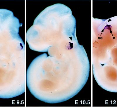
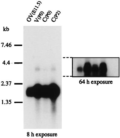
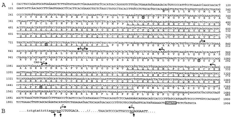
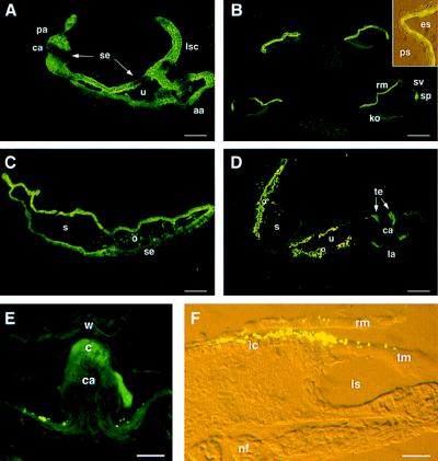
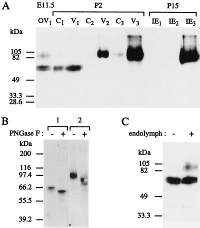
During the course of a study aimed at identifying inner ear-specific transcripts, a 1,906-bp murine cDNA predicted to encode a secreted 469-aa protein with two domains of homology with the secreted phospholipases A2 was isolated. This transcript is specifically expressed in the inner ear from embryonic day 9.5. The encoded 95-kDa glycoprotein is the major protein of the utricular and saccular otoconia and thus was named otoconin-95. By immunohistofluorescence, otoconin-95 also was detected in the cupulae of the semicircular canals and in previously undescribed transient granular structures of the cochlea. Otoconin-95 was found to be synthesized by various nonsensory cell types, but not by the supporting cells of the sensory epithelia, which produce the otoconial precursor vesicles. In addition, multiple isoforms generated by differential splicing were observed in different combinations during development. Based on the present results, we propose a model for the formation of the otoconia.


Figures

Figure 1
Figure 1
Expression of Ocn-95 at early embryonic stages. At E9.5–E10.5, the growing endolymphatic duct (arrowhead) and the dorsal part of the otic vesicle are strongly labeled. At E12, the endolymphatic duct, the forming semicircular canals (sc), and the cochlea (c) are stained. Magnification: ×25 (E9.5), ×17 (E10.5), and ×6 (E12).
Figure 2
Figure 2
Northern blot analysis of mouse poly(A)+ RNA at E11.5, P0, and P2. OV, otic vesicle; V, vestibule; C, cochlea.
Figure 3
Figure 3
(A) Ocn-95 cDNA and deduced amino acid sequences. The translation initiation codon is underlined, and a polyadenylation signal is boxed. The alternative regions (A, B, C, and CAG insertion) are indicated by broken arrows. The CAG insertion at position 1096 is predicted to result in the insertion of an alanine at position 298. Loss of A, (A + B), and C is predicted to result in in-frame deletions of 17 (G208-E224), 33 (G208-T240), and 34 (A297-P330) aa, respectively. Loss of (A+B) is also responsible for an amino acid substitution (K241D). Regions A and B are, according to the structure of PLA2L (28), encoded by two adjacent exons. The two sPLA2-like domains are underlined, and four potential N-glycosylation sites are circled. (B) Alternative splicing at the intron/region C junction. Lowercase and uppercase letters denote intronic and exonic sequences, respectively. The alternative region C is indicated between broken arrows. The three acceptor splice sites (canonical AG underlined) are highlighted by vertical arrows. Region C corresponds to the 5′ part of a 172-bp human exon (28) and its 3′ end fits well with an acceptor splice site (29). Amplification on mouse genomic DNA of a 132-bp fragment between T1100 and G1231 confirmed that the 102-bp region C corresponds to the 5′ part of a longer exon. Sequence analysis of the intron-region C junction revealed two overlapping acceptor splice sites. The use of the most 5′ one results in the CAG insertion at position 1096.
Figure 4
Figure 4
Distribution of otoconin-95 in the developing inner ear. (A) Section through the three ampullae and part of the utriculo-saccular complex at E14.5. (B) Four sections of the cochlear duct at P0. Note that cochlear cell differentiation proceeds with a base-to-apex gradient of maturation. (Inset) A detailed view of the two cell layers of the Reissner’s membrane. (C) Saccule at P0. (D) Vestibule at P4. (E) Posterior ampulla at P21. The punctate signals above the epithelium on each side of the cristae were not reproducible; they may correspond to displaced utricular otoconia. (F) Cochlea at P8. Inset and F micrographs were taken under both daylight and fluorescent light with Nomarski optics. The 046CP4 serum was used at 1:1,000 dilution. aa, anterior ampulla; c, cupula; ca, crista ampullaris; es, endolymphatic space; ic, interdental cells; is, inner sulcus; ko, Kölliker’s organ; la, lateral ampulla; lsc, lateral semicircular canal; nf, nerve fiber; o, otoconia; pa, posterior ampulla; ps, perilymphatic space; rm, Reissner’s membrane; s, saccule; se, sensory epithelium; sp, spiral prominence; sv, stria vascularis; te, transitory epithelium; tm, tectorial membrane; u, utricle; w, wall of the ampulla. (Scale bars: A, B, and D, 50 μm; C and E, 30 μm; F, 15 μm.)
Figure 5
Figure 5
Immunoblot analysis of otoconin-95. (A) Differential extraction of otoconin-95 isoforms. Soluble proteins were extracted from otic vesicle at E11.5 (OV1), cochlea (C1), and vestibule (V1) at P2, and inner ear (IE1) at P15. Proteins of the remaining insoluble fractions were sequentially extracted by sonication in the presence of 0.2% SDS (C2, V2, and IE2, respectively) and decalcification in the presence of 20% EDTA (C3, V3, and IE3, respectively). (B) N-glycosydase F sensitivity of the 65-kDa (lane 1) and 85- to 105-kDa (lane 2) forms. Fractions C1 and V2 were incubated either with (+) or without (−) N-glycosydase F (PNGase F). (C) Identification of the intracellular and secreted isoforms of otoconin-95. Proteins were extracted from E11.5 otic vesicles in which endolymph had been retained (+) or removed (−). Molecular mass standards are indicated on the left.
Figure 6
Figure 6
Protein content of the otoconial complex. Immunodetection of otoconin-95 (1/15 of the sample, lanes 1 and 2) and colloidal gold staining (14/15 of the sample, lanes 4 and 5) of proteins extracted from the otoconial membranes (lanes 1 and 4) and otoconia (lanes 2 and 5) of adult mice; lane 3, control lane for colloidal gold staining loaded without protein added. Molecular masses (in kDa) of the marker proteins in lane M are indicated on the right. Note that the bands, migrating between the 55.6- and 66.4-kDa markers, observed in each lane (including lane 3) are artifacts seen only in the presence of reducing agents.

References

    1. Carlström D. Biol Bull. 1963;125:441–463.
    1. Marmo F, Balsamo G, Franco E. Cell Tissue Res. 1983;233:35–43. - PubMed
    1. Pote K G, Ross M D. Comp Biochem Physiol. 1991;98:287–295. - PubMed
    1. Pote K G, Ross M D. J Ultrastruct Mol Struct Res. 1986;95:61–70. - PubMed
    1. Pote K G, Hauer III C R, Michel H, Shabanowitz J, Hunt D F, Kretsinger R H. Biochemistry. 1993;32:5017–5024. - PubMed

Publication types

MeSH terms

Associated data