

Paralogous mouse Hox genes, Hoxa9, Hoxb9, and Hoxd9, function together to control development of the mammary gland in response to pregnancy
- PMID: 9892669
- PMCID: PMC15172
- DOI: 10.1073/pnas.96.2.541
Paralogous mouse Hox genes, Hoxa9, Hoxb9, and Hoxd9, function together to control development of the mammary gland in response to pregnancy
Abstract
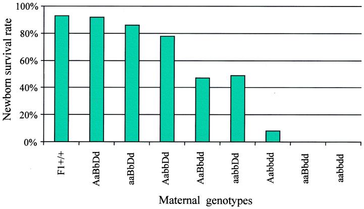
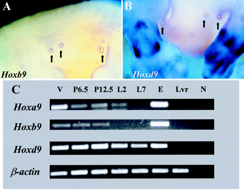
Although the role of Hox genes in patterning the mammalian body plan has been studied extensively during embryonic and fetal development, relatively little is known concerning Hox gene function in adult animals. Analysis of mice with mutant Hoxa9, Hoxb9, and Hoxd9 genes shows that these paralogous genes are required for mediating the expansion and/or differentiation of the mammary epithelium ductal system in response to pregnancy. Mothers with these three mutant genes cannot raise their own pups, but the pups can be rescued by fostering by wild-type mothers. Histologically, the mammary glands of the mutant mothers seem normal before pregnancy but do not develop properly in response to pregnancy and parturition. Hoxa9, Hoxb9, and Hoxd9 are expressed normally in adult mammary glands, suggesting a direct role for these genes in the development of mammary tissue after pregnancy. Because loss-of-function mutations in these Hox genes cause hypoplasia of the mammary gland after pregnancy, it may be productive to look for misexpression of these genes in mammary carcinomas.
Figures

Comment in
-
No milk today (my Hox have gone away).Proc Natl Acad Sci U S A. 1999 Jan 19;96(2):322-3. doi: 10.1073/pnas.96.2.322. Proc Natl Acad Sci U S A. 1999. PMID: 9892628 Free PMC article. No abstract available.
Similar articles
-
Key pathways regulated by HoxA9,10,11/HoxD9,10,11 during limb development.BMC Dev Biol. 2015 Jul 19;15:28. doi: 10.1186/s12861-015-0078-5. BMC Dev Biol. 2015. PMID: 26186931 Free PMC article.
-
Hox genes in normal and neoplastic mouse mammary gland.Cancer Res. 1994 Nov 15;54(22):5981-5. Cancer Res. 1994. PMID: 7954431
-
No milk today (my Hox have gone away).Proc Natl Acad Sci U S A. 1999 Jan 19;96(2):322-3. doi: 10.1073/pnas.96.2.322. Proc Natl Acad Sci U S A. 1999. PMID: 9892628 Free PMC article. No abstract available.
-
HOX genes function in Breast Cancer development.Biochim Biophys Acta Rev Cancer. 2020 Apr;1873(2):188358. doi: 10.1016/j.bbcan.2020.188358. Epub 2020 Mar 5. Biochim Biophys Acta Rev Cancer. 2020. PMID: 32147544 Review.
-
HOXA9 versus HOXB9; particular focus on their controversial role in tumor pathogenesis.J Appl Genet. 2024 Sep;65(3):473-492. doi: 10.1007/s13353-024-00868-x. Epub 2024 May 16. J Appl Genet. 2024. PMID: 38753266 Review.
Cited by
-
HOXC9 Induces Phenotypic Switching between Proliferation and Invasion in Breast Cancer Cells.J Cancer. 2016 Apr 10;7(7):768-73. doi: 10.7150/jca.13894. eCollection 2016. J Cancer. 2016. PMID: 27162534 Free PMC article.
-
Hox11 paralogous genes are essential for metanephric kidney induction.Genes Dev. 2002 Jun 1;16(11):1423-32. doi: 10.1101/gad.993302. Genes Dev. 2002. PMID: 12050119 Free PMC article.
-
HOXB9, a gene overexpressed in breast cancer, promotes tumorigenicity and lung metastasis.Proc Natl Acad Sci U S A. 2010 Jan 19;107(3):1100-5. doi: 10.1073/pnas.0912710107. Epub 2009 Dec 28. Proc Natl Acad Sci U S A. 2010. PMID: 20080567 Free PMC article.
-
Loss of HOXB3 correlates with the development of hormone receptor negative breast cancer.PeerJ. 2020 Nov 20;8:e10421. doi: 10.7717/peerj.10421. eCollection 2020. PeerJ. 2020. PMID: 33240685 Free PMC article.
-
Role of homeobox genes in the patterning, specification, and differentiation of ectodermal appendages in mammals.J Cell Physiol. 2008 Aug;216(2):337-46. doi: 10.1002/jcp.21491. J Cell Physiol. 2008. PMID: 18459147 Free PMC article. Review.
References
-
- Krumlauf R. Cell. 1994;78:191–201. - PubMed
-
- Maconochie M, Nonchev S, Morrison A, Krumlauf R. Annu Rev Genet. 1996;30:529–556. - PubMed
-
- Capecchi M R. In: Molecular and Cellular Aspects of Neural Development. Cowan W M, Jessell W M, Zipursky S L, editors. New York: Oxford Univ. Press; 1997. pp. 334–355.
-
- Capecchi M R. Cold Spring Harbor Symposia on Quantitative Biology. Vol. 62. Plainview, NY: Cold Spring Harbor Lab. Press; 1997. pp. 273–281. - PubMed
-
- Condie B G, Capecchi M R. Nature (London) 1994;370:304–307. - PubMed
Publication types
MeSH terms
Substances
LinkOut - more resources
Full Text Sources
Molecular Biology Databases
