. 1999 Jan 19;96(2):552-7.
doi: 10.1073/pnas.96.2.552.

MyoR: a muscle-restricted basic helix-loop-helix transcription factor that antagonizes the actions of MyoD

Affiliations

MyoR: a muscle-restricted basic helix-loop-helix transcription factor that antagonizes the actions of MyoD

J Lu et al. Proc Natl Acad Sci U S A. .

Abstract

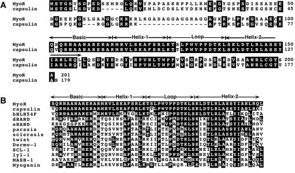
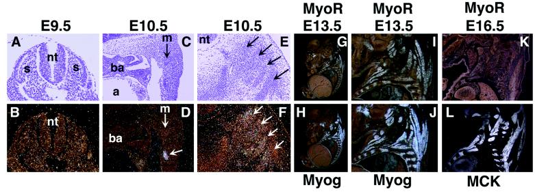
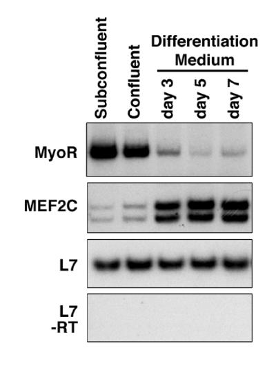
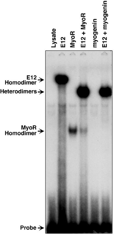
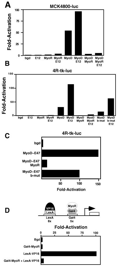
Skeletal muscle development is controlled by a family of muscle-specific basic helix-loop-helix (bHLH) transcription factors that activate muscle genes by binding E-boxes (CANNTG) as heterodimers with ubiquitous bHLH proteins, called E proteins. Myogenic bHLH factors are expressed in proliferating undifferentiated myoblasts, but they do not initiate myogenesis until myoblasts exit the cell cycle. We describe a bHLH protein, MyoR (for myogenic repressor), that is expressed in undifferentiated myoblasts in culture and is down-regulated during differentiation. MyoR is also expressed specifically in the skeletal muscle lineage between days 10.5 and 16.5 of mouse embryogenesis and down-regulated thereafter during the period of secondary myogenesis. MyoR forms heterodimers with E proteins that bind the same DNA sequence as myogenic bHLH/E protein heterodimers, but MyoR acts as a potent transcriptional repressor that blocks myogenesis and activation of E-box-dependent muscle genes. These results suggest a role for MyoR as a lineage-restricted transcriptional repressor of the muscle differentiation program.


Figures

Figure 1
Figure 1
Conceptual ORF of MyoR and homology with other bHLH proteins. (A) The deduced ORF of MyoR and amino acid alignment with capsulin is shown. (B) Homology between the bHLH region of MyoR and other bHLH proteins.
Figure 2
Figure 2
Detection of MyoR and myogenin expression in developing skeletal muscle by in situ hybridization. Transcripts for MyoR were detected in transverse sections of E9.5 (B) and E10.5 (F) and in sagittal sections of E10.5 (D), E13.5 (G and I), and E16.5 (K) embryos. Expression of myogenin at E13.5 is illustrated in sagittal sections (H and J). Transcripts for MCK are shown in sagittal section of an E16.5 embryo (L). I and J show enlarged views of G and H, respectively. At E16.5, MyoR expression is down-regulated in contrast to the abundant expression of MCK. a, Atrium; ba, branchial arch; m, myotome; nt, neural tube; s, somite. The arrow in D shows a cluster of MyoR-expressing cells in body wall ventrolateral to the myotome (see text). Arrows in E and F mark differentiating axial skeletal muscle. (A and B, ×60; CF, ×30; G and H, ×4; I and J, ×8; and K and L, ×5.)
Figure 3
Figure 3
Expression of MyoR transcripts during differentiation of C2 cells. Total RNA was isolated from C2 cells maintained at subconfluent or confluent densities in growth medium or after transfer to differentiation medium for 3, 5, and 7 days. Transcripts for MyoR, MEF2C, and L7 were measured by semiquantitative reverse transcriptase–PCR. MyoR and L7 yield single PCR products of the predicted size, and MEF2C yields two products reflecting alternative splicing. In the absence of reverse transcriptase (-RT), no products were observed for L7 (Bottom), MyoR, or MEF2C (not shown).
Figure 4
Figure 4
Binding of MyoR/E12 heterodimers to DNA. E12, MyoR, and/or myogenin were transcribed and translated separately or together, as indicated, using a coupled in vitro transcription/translation system. In vitro translation products were used in gel mobility-shift assays with an end-labeled double-stranded oligonucleotide probe corresponding to the MCK right E-box. In the presence of lysate alone, no binding activity was observed.
Figure 5
Figure 5
MyoR inhibits transcription. 10T½ cells were transiently transfected with MCK4800-luc (A) or 4R-tk-luc (B and C) and expression vectors encoding the indicated bHLH proteins. The background level of expression of the reporters without cotransfected bHLH expression vectors (bgd) was assigned a value of 1. (D) COS cells were transiently transfected with L8G5-luc reporter and expression vectors encoding LexA-VP16 and GAL4-MyoR, as indicated. The amount of DNA in each transfection was kept constant by addition of parent vector pM1. GAL4-MyoR resulted in greater than a 20-fold reduction in reporter gene expression in the presence of LexA-VP16. Assays were performed three times with comparable results. Luciferase activity was normalized to activity of β-galactosidase, obtained by cotransfection of RSV-lacZ as an internal control.

Similar articles

Cited by

References

    1. Yun S, Wold B J. Curr Opin Cell Biol. 1996;8:877–889. - PubMed
    1. Molkentin J, Olson E N. Curr Opin Genet Dev. 1996;6:445–453. - PubMed
    1. Rudnicki M A, Schnegelsberg P N J, Stead R H, Braun T, Arnold H H, Jaenisch R. Cell. 1993;71:1351–1359. - PubMed
    1. Hasty P, Bradley A, Morris J H, Venuti J M, Olson E N, Klein W H. Nature (London) 1993;364:501–506. - PubMed
    1. Nabeshima Y K, Hanaoka K, Hayasaka M, Esumi S, Li S, Nonaka I, Nabeshima Y. Nature (London) 1993;364:532–535. - PubMed

Publication types

MeSH terms

Associated data