

Isolation and expression of a Pax-6 gene in the regenerating and intact Planarian Dugesia(G)tigrina
- PMID: 9892672
- PMCID: PMC15175
- DOI: 10.1073/pnas.96.2.558
Isolation and expression of a Pax-6 gene in the regenerating and intact Planarian Dugesia(G)tigrina
Abstract
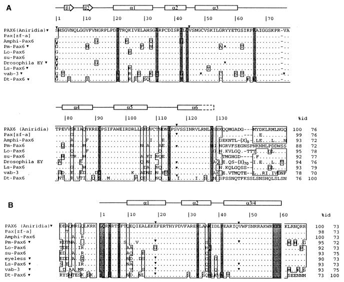
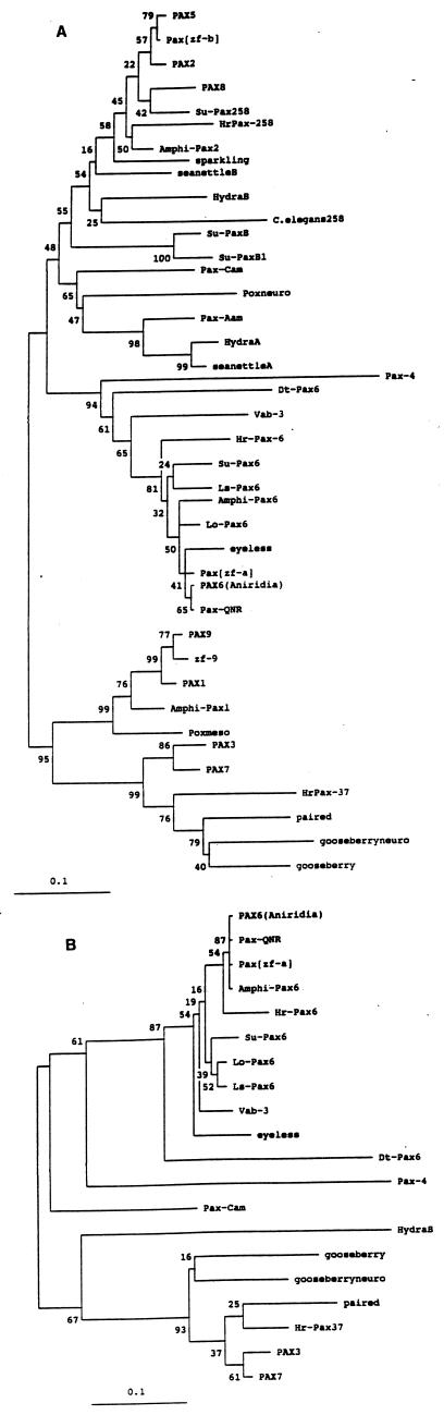
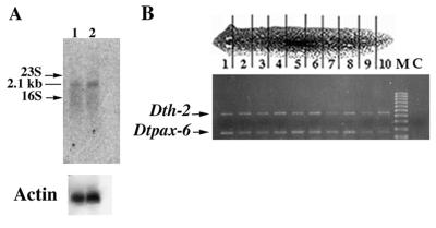
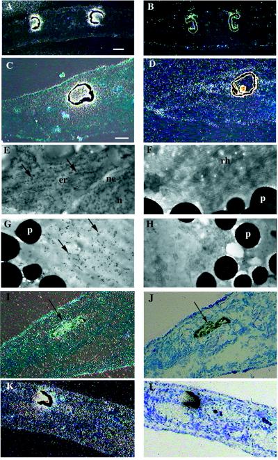
The Pax-6 gene encodes a transcription factor containing both a paired and a homeodomain and is highly conserved among Metazoa. In both vertebrates and invertebrates, Pax-6 is required for eye morphogenesis, development of parts of the central nervous system, and, in some phyla, for the development of olfactory sense organs. Ectopic expression of Pax-6 from insects, mammals, cephalopods, and ascidians induces ectopic eyes in Drosophila, suggesting that Pax-6 may be a universal master control gene for eye morphogenesis. Platyhelminthes are an ancient phylum, originating from the base of spiralian protostomes, that bear primitive eyes, consisting of a group of rhabdomeric photoreceptor cells enclosed in a cup of pigment cells. The analysis of Pax-6 and its expression pattern should provide insights into the ancestral function of Pax-6 in eye morphogenesis. We have identified the Pax-6 gene of the planarian Dugesia(G)tigrina (Platyhelminthes; Turbellaria; Tricladida). This gene shares significant sequence identity and conserved genomic organization with Pax-6 proteins from other phyla. Phylogenetic analysis indicates that it clusters with the other Pax-6 genes, but in the most basal position. DtPax-6 is expressed as a single transcript in both regenerating and fully grown eyes, and electron microscopy studies show strong expression in the perykarion of both photoreceptor and pigment cells. Very low levels of expression also are detectable in other body regions. Because a bona fide Pax-6 homolog so far has not been detected in diploblastic animals, we speculate that Pax-6 may be typical for triploblasts and that the appearance of additional Pax genes may have coincided with increasingly complex body plans.
Figures
Similar articles
-
Conservation of Pax-6 in a lower chordate, the ascidian Phallusia mammillata.Development. 1997 Feb;124(4):817-25. doi: 10.1242/dev.124.4.817. Development. 1997. PMID: 9043063
-
Isolation of a Pax-6 homolog from the ribbonworm Lineus sanguineus.Proc Natl Acad Sci U S A. 1996 Apr 2;93(7):2658-63. doi: 10.1073/pnas.93.7.2658. Proc Natl Acad Sci U S A. 1996. PMID: 8610097 Free PMC article.
-
Isolation and developmental expression of the amphioxus Pax-6 gene (AmphiPax-6): insights into eye and photoreceptor evolution.Development. 1998 Jul;125(14):2701-10. doi: 10.1242/dev.125.14.2701. Development. 1998. PMID: 9636084
-
Pax 6: mastering eye morphogenesis and eye evolution.Trends Genet. 1999 Sep;15(9):371-7. doi: 10.1016/s0168-9525(99)01776-x. Trends Genet. 1999. PMID: 10461206 Review.
-
The master control gene for morphogenesis and evolution of the eye.Genes Cells. 1996 Jan;1(1):11-5. doi: 10.1046/j.1365-2443.1996.11011.x. Genes Cells. 1996. PMID: 9078363 Review.
Cited by
-
Cognitive Stimulation Induces Differential Gene Expression in Octopus vulgaris: The Key Role of Protocadherins.Biology (Basel). 2020 Jul 30;9(8):196. doi: 10.3390/biology9080196. Biology (Basel). 2020. PMID: 32751499 Free PMC article.
-
The Pax gene family: Highlights from cephalopods.PLoS One. 2017 Mar 2;12(3):e0172719. doi: 10.1371/journal.pone.0172719. eCollection 2017. PLoS One. 2017. PMID: 28253300 Free PMC article.
-
Dicyema Pax6 and Zic: tool-kit genes in a highly simplified bilaterian.BMC Evol Biol. 2007 Oct 25;7:201. doi: 10.1186/1471-2148-7-201. BMC Evol Biol. 2007. PMID: 17961212 Free PMC article.
-
Searching for the prototypic eye genetic network: Sine oculis is essential for eye regeneration in planarians.Proc Natl Acad Sci U S A. 2000 Apr 25;97(9):4525-9. doi: 10.1073/pnas.97.9.4525. Proc Natl Acad Sci U S A. 2000. PMID: 10781056 Free PMC article.
-
Differential expression of retinal determination genes in the principal and secondary eyes of Cupiennius salei Keyserling (1877).Evodevo. 2015 Apr 28;6:16. doi: 10.1186/s13227-015-0010-x. eCollection 2015. Evodevo. 2015. PMID: 26034575 Free PMC article.
References
-
- Valentine J W, Erwin D H, Jablonski D. Developmental Biology. 1996;173:373–381. - PubMed
-
- Slack J M W, Holland P W H, Graham C F. Nature (London) 1993;361:490–492. - PubMed
-
- Bopp D, Burri M, Baumgartner S, Frigerio G, Noll M. Cell. 1986;47:1033–1040. - PubMed
-
- Callaerts P, Halder G, Gehring W J. Annu Rev Neurosci. 1997;20:483–532. - PubMed
Publication types
MeSH terms
Substances
Associated data
- Actions
LinkOut - more resources
Full Text Sources
