

Generation of native bovine mAbs by phage display
- PMID: 9892686
- PMCID: PMC15189
- DOI: 10.1073/pnas.96.2.640
Generation of native bovine mAbs by phage display
Abstract
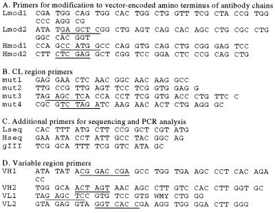
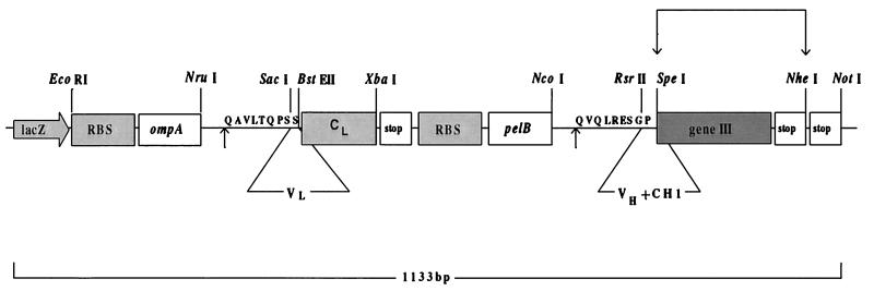
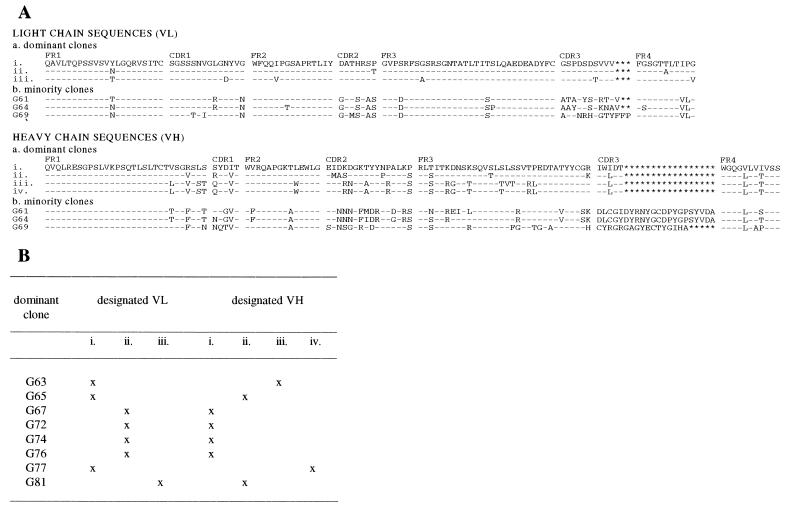
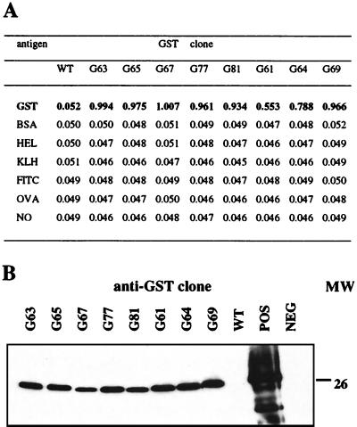
Modeling of disease pathogenesis and immunity often is carried out in large animals that are natural targets for pathogens of human or economic relevance. Although murine mAbs are a valuable tool in identifying certain host/pathogen interactions, progress in comparative immunology would be enhanced by the use of mAbs isolated from the host species. Such antibodies would reflect an authentic host immune response to infection or vaccination, and as they are host derived, would allow the application of in vivo experiments that previously have been unrealizable in large animals because of induction of an antispecies immune response. The advent of antibody phage display technology provides a way of producing host-derived mAbs in animals where the molecular genetics of Ig formation are known. Exploiting recent advances in the molecular immunology of cattle, we report here the design of an optimized phage display vector, pComBov, for the construction of combinatorial libraries of bovine Ig antigen-binding fragments (Fab) of native sequence. By using this system, we initially have generated and characterized a panel of bovine mAbs against a model antigen glutathione S-transferase. The isolated mAbs showed features typical of bovine Igs and recognized glutathione S-transferase with high specificity in ELISA and by Western blotting. The pComBov expression system can be readily adapted for the preparation of libraries from related ruminant species and advances the use of monoclonal reagents derived in this way for comparative studies in animals of economic importance.
Figures
Similar articles
-
Conversion of murine Fabs isolated from a combinatorial phage display library to full length immunoglobulins.J Immunol Methods. 1995 Aug 18;184(2):177-86. doi: 10.1016/0022-1759(95)00086-p. J Immunol Methods. 1995. PMID: 7658022
-
An improved phage display antibody cloning system using newly designed PCR primers optimized for Pfu DNA polymerase.J Biochem. 1995 Jun;117(6):1218-27. doi: 10.1093/oxfordjournals.jbchem.a124847. J Biochem. 1995. PMID: 7490263
-
Human monoclonal recombinant Fabs specific for HCV antigens obtained by repertoire cloning in phage display combinatorial vectors.Res Virol. 1997 Mar-Apr;148(2):165-9. doi: 10.1016/s0923-2516(97)89904-9. Res Virol. 1997. PMID: 9108620
-
Molecular considerations for development of phage antibody libraries.J Drug Target. 2012 Apr;20(3):195-208. doi: 10.3109/1061186X.2011.611517. Epub 2011 Sep 27. J Drug Target. 2012. PMID: 21950316 Review.
-
Monoclonal Antibodies and Antibody Like Fragments Derived from Immunised Phage Display Libraries.Adv Exp Med Biol. 2017;1053:99-117. doi: 10.1007/978-3-319-72077-7_6. Adv Exp Med Biol. 2017. PMID: 29549637 Free PMC article. Review.
Cited by
-
Wide screening of phage-displayed libraries identifies immune targets in planta.PLoS One. 2013;8(1):e54654. doi: 10.1371/journal.pone.0054654. Epub 2013 Jan 25. PLoS One. 2013. PMID: 23372747 Free PMC article.
-
A large semi-synthetic single-chain Fv phage display library based on chicken immunoglobulin genes.BMC Biotechnol. 2004 Apr 1;4:6. doi: 10.1186/1472-6750-4-6. BMC Biotechnol. 2004. PMID: 15059288 Free PMC article.
-
Natural and man-made V-gene repertoires for antibody discovery.Front Immunol. 2012 Nov 15;3:342. doi: 10.3389/fimmu.2012.00342. eCollection 2012. Front Immunol. 2012. PMID: 23162556 Free PMC article.
-
Phage Display Screening of Bovine Antibodies to Foot-and-Mouth Disease Virus and Their Application in a Competitive ELISA for Serodiagnosis.Int J Mol Sci. 2021 Apr 21;22(9):4328. doi: 10.3390/ijms22094328. Int J Mol Sci. 2021. PMID: 33919326 Free PMC article.
-
A Broad Role for Cysteines in Bovine Antibody Diversity.Immunohorizons. 2019 Oct 16;3(10):478-487. doi: 10.4049/immunohorizons.1900058. Immunohorizons. 2019. PMID: 31619454 Free PMC article.
References
Publication types
MeSH terms
Substances
Associated data
- Actions
- Actions
- Actions
- Actions
- Actions
- Actions
- Actions
- Actions
- Actions
- Actions
- Actions
- Actions
- Actions
LinkOut - more resources
Full Text Sources
Other Literature Sources
