. 1999 Jan 19;96(2):657-62.
doi: 10.1073/pnas.96.2.657.

Nitric oxide synthase in cardiac sarcoplasmic reticulum

Affiliations

Nitric oxide synthase in cardiac sarcoplasmic reticulum

K Y Xu et al. Proc Natl Acad Sci U S A. .

Abstract

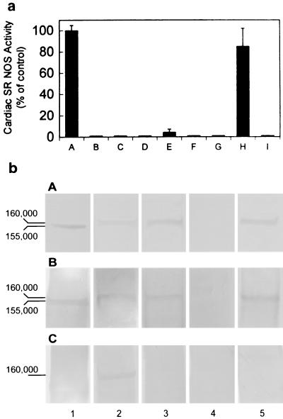
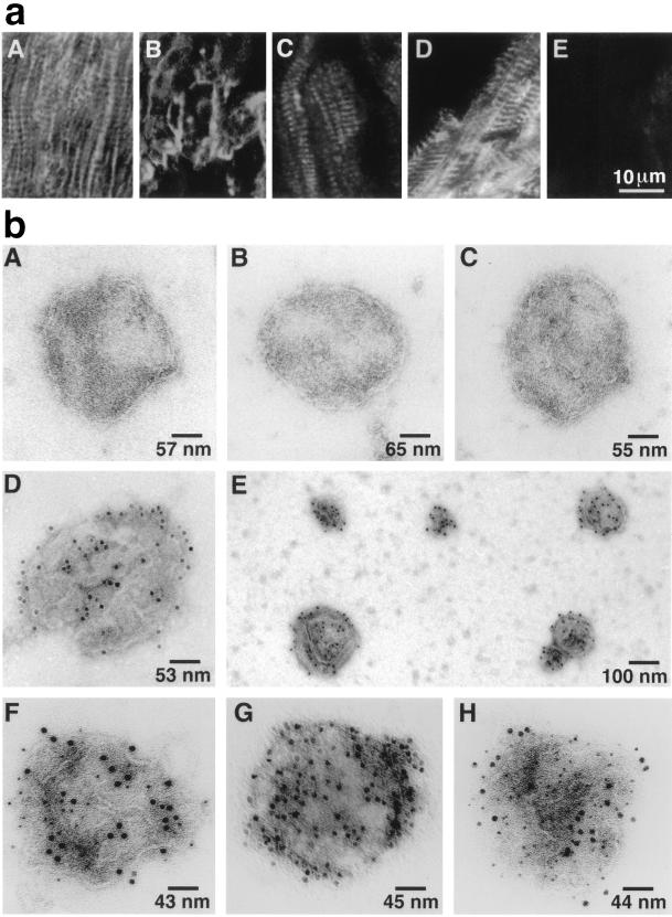
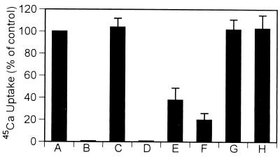
NO. is a free radical that modulates heart function and metabolism. We report that a neuronal-type NO synthase (NOS) is located on cardiac sarcoplasmic reticulum (SR) membrane vesicles and that endogenous NO. produced by SR-associated NOS inhibits SR Ca2+ uptake. Ca2+-dependent biochemical conversion of L-arginine to L-citrulline was observed from isolated rabbit cardiac SR vesicles in the presence of NOS substrates and cofactors. Endogenous NO. was generated from the vesicles and detected by electron paramagnetic resonance spin-trapping measurements. Immunoelectron microscopy demonstrated labeling of cardiac SR vesicles by using anti-neuronal NOS (nNOS), but not anti-endothelial NOS (eNOS) or anti-inducible NOS (iNOS) antibodies, whereas skeletal muscle SR vesicles had no nNOS immunoreactivity. The nNOS immunoreactivity also displayed a pattern consistent with SR localization in confocal micrographs of sections of human myocardium. Western blotting demonstrated that cardiac SR NOS is larger than brain NOS (160 vs. 155 kDa). No immunodetection was observed in cardiac SR vesicles from nNOS knockout mice or with an anti-nNOS mu antibody, suggesting the possibility of a new nNOS-type isoform. 45Ca uptake by cardiac SR vesicles, catalyzed by Ca2+-ATPase, was inhibited by NO. produced endogenously from cardiac SR NOS, and 7-nitroindazole, a selective nNOS inhibitor, completely prevented this inhibition. These results suggest that a cardiac muscle nNOS isoform is located on SR of cardiac myocytes, where it may respond to intracellular Ca2+ concentration and modulate SR Ca2+ ion active transport in the heart.


Figures

Figure 1
Figure 1
(a) NO synthase activity was found in isolated cardiac SR vesicles. Lane A: control, in the presence of NOS substrates and cofactors: Ca2+, CaM, NADPH, FMN, FAD, [3H]arginine, and BH4; lane B: in the presence of NOS substrates without vesicles; lane C: boiled vesicles + NOS substrates and cofactors; lane D: in the absence of Ca2+; lane E: with EGTA and NOS substrates and cofactors; lane F: with 10 μM 7-NI; lane G: with 60 μM TRIM; lane H: with 10 nM AMT; and lane I: iNOS in the presence of 10 nM AMT. These results demonstrate that SR NOS activity is Ca2+ dependent and is sensitive to nNOS selective inhibitors, indicating the existence of NOS in cardiac SR vesicles and the specificity of a neuronal-type NOS enzymatic activity. The data represent a mean of six independent experiments. (b) Western blot analysis of cardiac SR NOS. Cardiac SR vesicles were fractionated by electrophoresis through a 7.5% SDS-polyacrylamide gel and transferred to nitrocellulose. Incubated with Affinity BioReagents anti-nNOS antibody (bA), with anti-nNOSα antibody (bB), and with anti-nNOSμ antibody (bC). Column 1: rat brain homogenate; 2: mice heart homogenate; 3: mice cardiac SR vesicles; 4: nNOS knockout mice cardiac SR vesicles; and 5: rabbit cardiac SR vesicles. All of the samples were 30 μg (total protein) per lane. Both rabbit and mice cardiac SR NOS were detected by anti-nNOS and anti-αnNOS antibodies. The results also show that the denatured molecular masses of rat brain NOS and cardiac SR NOS were ≈155 and 160 kDa, respectively.
Figure 2
Figure 2
EPR spectra of NO formation from cardiac SR vesicles in the presence of a Fe(MGD)2 complex under various conditions for 60 min at room temperature. Fe(MGD)2 only (A); + SOD in condition A (B); + a mixture of NOS substrates and cofactors in condition B (C); + cardiac vesicles in condition C (D); + 7-NI in condition D (E); and + d-arginine in the presence of vesicles and cofactors (F) (see Methods for the final concentrations of the reaction components). These results show that 7-NI sensitive, endogenous NO (a triplet spectrum) is generated by the isolated cardiac SR vesicles. The data are presented from one of four similar independent experiments in each case.
Figure 3
Figure 3
(a) Immunofluorescent localization of cardiomyocyte antigens with mAbs on cryostat sections of human ventricular myocardium by laser-scanning confocal microscopy. (aA) nNOS indirect immunofluorescent labeling (IIL) shows a linear longitudinal and transverse, striated pattern of labeling; Na+,K+-ATPase IIL demonstrates a distinctly different sarcolemma pattern of labeling of the human heart section (aB); RyR IIL (aC) and SERCA2 IIL (aD) have a similar pattern of labeling as NOS IIL suggesting SR localization of NOS rather than exclusive sarcolemma localization in cardiac muscle; an irrelevant anti-viral protein mAb (aE) used at the same concentration as the other primary mAbs, has only low levels of background fluorescence. (b) Single immunogold labeling. SR vesicles were incubated with specific primary antibodies and a secondary antibody conjugated to 12 nm of colloidalgold. Control vesicles in the absence of primary antibody (bA); with anti-eNOS (bB); with anti-iNOS (bC); and single vesicle (bD) and a group of vesicles (bE) with anti-nNOS. Electron photomicrographs show that only nNOS is broadly dispersed on the surface of cardiac muscle SR membrane vesicles. Double immunogold labeling: Ca2+-ATPase (6 nm gold particles) and nNOS (12 nm) (bF); Na+, K+-ATPase (6 nm) and nNOS (12 nm) (bG); and phospholamban (6 nm) and nNOS (12 nm) (bH). Each vesicle represents hundreds of similar labeled or unlabeled vesicles in each condition. The results show that cardiac SR Ca2+-ATPase and phospholamban coexist with a SR neuronal-type NOS. Absence of Na+,K+-ATPase on the SR vesicles further demonstrates the vesicles represent SR rather than sarcolemma.
Figure 4
Figure 4
Effect of endogenous NO on cardiac SR 45Ca uptake under various conditions. Vesicles in the absence of NOS substrates, cofactors, and SOD (A); −SOD + thapsigargin (B) (Tg, a specific inhibitor of Ca2+-ATPase); vesicles + SOD (C); + SOD + Tg (D); condition C in the presence of NOS substrates and cofactors for 30 min (E), or for 60 min (F) (30 min at 23°C and 30 min at 37°C); 60-min incubation (same as F) in the presence of the NOS inhibitor 7-NI (10 μM) (G); d-arginine replaced l-arginine in condition F (H). Data represent the mean ± SD from six independent experiments by using 0.6 mg/ml cardiac SR vesicles. The concentration of SOD was 300 units/ml in all reaction mixtures. 45Ca uptake is inhibited by endogenously produced NO. The results suggest that cardiac NOS regulates SR 45Ca uptake by directly modifying Ca2+-ATPase function through endogenous NO.

Similar articles

Cited by

References

    1. Nakane M, Schmidt H H, Pollock J S, Forstermann U, Murad F. FEBS Lett. 1993;316:175–180. - PubMed
    1. Kobzik L, Reid M B, Bredt D S, Stamler J S. Nature (London) 1994;372:546–548. - PubMed
    1. Brenman J E, Chao D S, Xia H, Aldape K, Bredt D S. Cell. 1995;82:743–752. - PubMed
    1. Silvagno F, Xia H, Bredt D S. J Biol Chem. 1996;271:11204–11208. - PubMed
    1. Kelly R A, Balligad J L, Smith T W. Circ Res. 1996;79:363–380. - PubMed

Publication types

MeSH terms

LinkOut - more resources