. 1999 Jan 19;96(2):721-5.
doi: 10.1073/pnas.96.2.721.

Purification of serine racemase: biosynthesis of the neuromodulator D-serine

Affiliations

Purification of serine racemase: biosynthesis of the neuromodulator D-serine

H Wolosker et al. Proc Natl Acad Sci U S A. .

Abstract

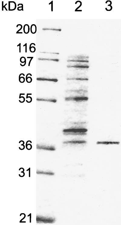
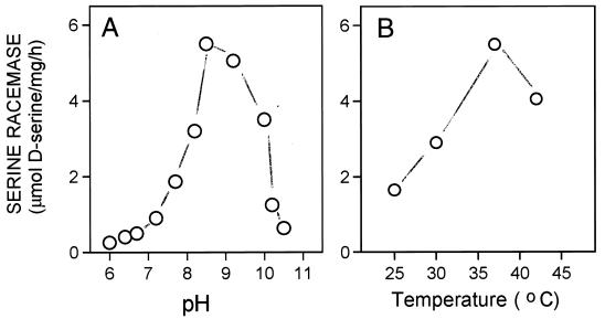
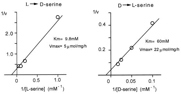
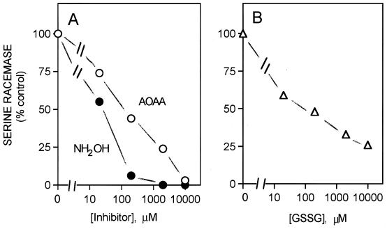
High levels of D-serine occur in mammalian brain, where it appears to be an endogenous ligand of the glycine site of N-methyl-D-aspartate receptors. In glial cultures of rat cerebral cortex, D-serine is enriched in type II astrocytes and is released upon stimulation with agonists of non-N-methyl-D-aspartate glutamate receptors. The high levels of D-serine in discrete areas of rat brain imply the existence of a biosynthetic pathway. We have purified from rat brain a soluble enzyme that catalyzes the direct racemization of L-serine to D-serine. Purified serine racemase has a molecular mass of 37 kDa and requires pyridoxal 5'-phosphate for its activity. The enzyme is highly selective toward L-serine, failing to racemize any other amino acid tested. Properties such as pH optimum, Km values, and the requirement for pyridoxal phosphate resemble those of bacterial racemases, suggesting that the biosynthetic pathway for D-amino acids is conserved from bacteria to mammalian brain.


Figures

Figure 1
Figure 1
SDS/PAGE analysis of purified serine racemase. A 12% polyacrylamide gel was stained with Coomassie blue. Lane 1, molecular mass markers: myosin (200 kDa), β-galactosidase (116.7 kDa), phosphorylase b (97.4 kDa), BSA (66.3 kDa), glutamic dehydrogenase (55.4 kDa), lactate dehydrogenase (36.5 kDa), carbonic anhydrase (31 kDa), and trypsin inhibitor (21.5 kDa). Lane 2, Mono Q column eluate containing 1 μg protein. Lane 3, hydroxyapatite column eluate containing 0.5 μg purified protein. Silver staining of the purified preparation showed no additional bands.
Figure 2
Figure 2
pH and temperature dependence of racemase activity. (A) Racemase activity was assayed at 37°C in media containing 50 mM Mes-Tris (pH 6.0–6.5), 50 mM Tris⋅HCl (pH 6.8–8.8) of 50 mM CAPS-NaOH (pH 9–10.5), 20 mM l-serine, 100 μg/ml purified enzyme, 1 mM EDTA, 2 mM DTT, and 15 μM PLP. (B) Racemase activity was performed at different temperatures in a medium containing 50 mM Tris⋅HCl, pH 8.0/20 mM l-serine, 100 μg/ml purified enzyme, 1 mM EDTA, 2 mM DTT, and 15 μM PLP. The reaction was stopped after 4 h and analyzed both by chemiluminescence and HPLC assay. The experiment was replicated three times by using different preparations with similar results.
Figure 3
Figure 3
Kinetic parameters of racemization reaction. Initial rate of racemase activity was measured at 37°C in medium containing 50 mM Tris⋅HCl, pH 8.0, 35 μg/ml purified enzyme, 1 mM EDTA, 2 mM DTT, and 15 μM PLP and different concentrations of either l- or d- serine. The reaction was stopped after 2 h when less than 10% substrate was consumed. Values for Km and Vmax were calculated by using the Michaelis–Menten equation. The values are representative of three experiments with different enzyme preparations.
Figure 4
Figure 4
Inhibition of serine racemase by PLP inhibitors and sulfhydryl oxidation. (A) Enzyme activity was monitored at 37°C in a medium containing 50 mM Tris⋅HCl, pH 8.0/20 mM l-serine, 100 μg/ml purified enzyme, 1 mM EDTA, 2 mM DTT, and 10 μM PLP and different concentrations of either AOAA (○) or hydroxylamine (•). (B) Reaction medium and conditions were as described in A, except that DTT was omitted from the last step of the enzyme preparation. The enzyme was preincubated for 10 min in the presence of different concentrations of oxidized glutathione (GSSG).
Figure 5
Figure 5
Absorption spectra of purified serine racemase. Purified enzyme (70 μg/ml) was preincubated for 10 min in medium containing 10 mM KPi (pH 7.2), 2 mM DTT, 1 mM EDTA, and 10 μM PLP, either in the absence (Control) or in the presence of 1 mM AOAA. The distinct peaks of absorbance at 420 and 340 nm were not observed in the presence of buffer alone or when BSA was used instead of serine racemase.

References

    1. Corrigan J J. Science. 1969;164:142–149. - PubMed
    1. Corrigan J J, Srinivasan N G. Biochemistry. 1966;5:1185–1190. - PubMed
    1. Nagata Y, Konno R, Yasumura Y, Akino T. Biochem J. 1989;257:291–292. - PMC - PubMed
    1. Hashimoto A, Nishikawa T, Hayashi T, Fujii N, Harada K, Oka T, Takahashi K. FEBS Lett. 1992;296:33–36. - PubMed
    1. Hashimoto A, Kumashiro S, Nishikawa T, Oka T, Takahashi K, Mito T, Takashima S, Doi N, Mizutani Y, Yamazaki T, Kaneko T, Ootomo E. J Neurochem. 1993;61:783–786. - PubMed

Publication types

LinkOut - more resources