. 1999 Jan 19;96(2):748-53.
doi: 10.1073/pnas.96.2.748.

Orexins, orexigenic hypothalamic peptides, interact with autonomic, neuroendocrine and neuroregulatory systems

Affiliations

Orexins, orexigenic hypothalamic peptides, interact with autonomic, neuroendocrine and neuroregulatory systems

Y Date et al. Proc Natl Acad Sci U S A. .

Abstract

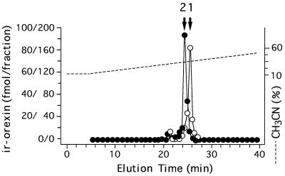
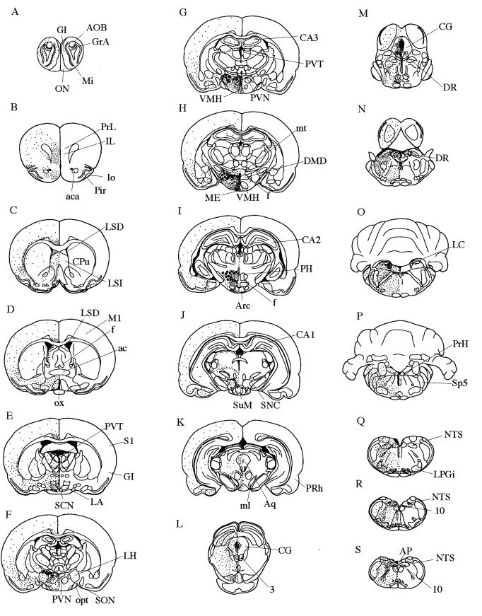
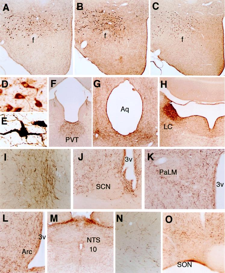
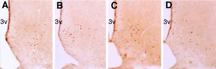
We determined the immunohistochemical distributions of orexin-A and orexin-B, hypothalamic peptides that function in the regulation of feeding behavior and energy homeostasis. Orexin-A and -B neurons were restricted to the lateral and posterior hypothalamus, whereas both orexin-A and -B nerve fibers projected widely into the olfactory bulb, cerebral cortex, thalamus, hypothalamus, and brainstem. Dense populations of orexin-containing fibers were present in the paraventricular thalamic nucleus, central gray, raphe nuclei, and locus coeruleus. Moderate numbers of these fibers were found in the olfactory bulb, insular, infralimbic and prelimbic cortex, amygdala, ventral, and dorsolateral parts of the suprachiasmatic nucleus, paraventricular nucleus except the lateral magnocellular division, arcuate nucleus, supramammillary nucleus, nucleus of the solitary tract, and dorsal motor nucleus of the vagus. Small numbers of orexin fibers were present in the perirhinal, motor and sensory cortex, hippocampus, and supraoptic nucleus, and a very small number in the lateral magnocellular division of the paraventricular nucleus. Intracerebroventricular injections of orexins induced c-fos expression in the paraventricular thalamic nucleus, locus coeruleus, arcuate nucleus, central gray, raphe nuclei, nucleus of the solitary tract, dorsal motor nucleus of the vagus, suprachiasmatic nucleus, supraoptic nucleus, and paraventricular nucleus except the lateral magnocellular division. The unique neuronal distribution of orexins and their functional activation of neural circuits suggest specific complex roles of the peptides in autonomic and neuroendocrine control.


Figures

Figure 1
Figure 1
Representative RP-HPLC profile of orexin-A (○) and -B (•) immunoreactivities in rat hypothalamus. Orexin-A content is represented by the left scale and orexin-B content by the right scale. Fraction volumes are 0.5 ml. Arrows indicate the elution positions of orexin-A (1) and orexin-B (2).
Figure 2
Figure 2
Localization of orexin-A and -B from rostal (A) to caudal (S) in rat brain. Orexin neurons: large filled circles. Orexin nerve fibers: small dots. 3, oculomotor nucleus; 10, dorsal motor nucleus of vagus; ac, anterior commissure; aca, anterior commissure, anterior part; AOB, accessory olfactory bulb; AP, area postrema; Aq, aqueduct; Arc, arcuate hypothalamic nucleus; CA1–3, fields CA1–3 of hippocampus; CG, central gray; CPu, caudate putamen; DMD, dorsomedial hypothalamic nucleus, dorsal part; DR, dorsal raphe nucleus; f, fornix; GI, granular insular cortex; Gl, glomerular layer of the olfactory bulb; GrA, granule cell layer of the accessory olfactory bulb; IL, infralimbic cortex; LA, lateroanterior hypothalamic nucleus; LC, locus coeruleus; LH, lateral hypothalamus; lo, lateral olfactory tract; LPGi, lateral paragigantocellular nucleus; LSD, lateral septal nucleus, dorsal part; LSI, lateral septal nucleus, intermediate part; M1, primary motor cortex; ME, median eminence; Mi, mitral cell layer of the olfactory bulb; ml, medial lemniscus; mt, mammillothalamic tract; NTS, nucleus of the solitary tract; ON, olfactory nerve layer; opt, optic tract; ox, optic chiasm; PH, posterior hypothalamus; Pir, piriform cortex; PRh, perirhinal cortex; PrH, prepositus hypoglossal nucleus; PrL, prelimbic cortex; PVN, paraventricular hypothalamic nucleus; PVT, paraventricular thalamic nucleus; S1, primary somatosensory cortex; SCN, suprachiasmatic nucleus; SNC, substantia nigra, compact part; SON, supraoptic nucleus; Sp5, spinal trigeminal nucleus; SuM, supramammillary nucleus; VMH, ventromedial hypothalamic nucleus.
Figure 3
Figure 3
Immunohistochemical localizations of orexin-A, orexin-A[17–33], and orexin-B. Neurons immunoreactive for orexin-A (A) and orexin-A[17–33] (B) are present in the perifornical region of the LH and posterior hypothalamic area. Orexin-B-immunoreactive neurons are present in the same areas (C). Orexin-A immunoreactive neurons in the LH at high magnification. Control (D), colchicine-treated rats (E). Orexin-A nerve fibers in the PVT (F), central gray and dorsal raphe nucleus (G), LC (H), infralimbic cortex (I), SCN (J), paraventricular nucleus (K), arcuate nucleus (Arc) (L), NTS, and dorsal motor nucleus of the vagus (10) (M), motor cortex (N), or SON (O). Abbreviations: f, fornix; Aq, aqueduct; 3v, third ventricle; PaLM, lateral magnocellular division of the paraventricular nucleus. Original magnification (AC, FH ×30, D, E ×280, IL, N, O ×70, M ×35.)
Figure 4
Figure 4
Fos immunoreactivity in the arcuate nucleus of the (A) 30-μg orexin-A-, (B) 3-μg orexin-A-, (C) 30-μg orexin-B-, and (D) 3- μg orexin-B-injected rats. 3v, third ventricle. Original magnification (AD ×70.)
Figure 5
Figure 5
Fos immunoreactivity in the arcuate nucleus (A and I), paraventricular thalamic nucleus (B and J), locus coeruleus (C and K), central gray and dorsal raphe nucleus (D and L), NTS and dorsal motor nucleus of the vagus (10) (E), suprachiasmatic nucleus (F), supraoptic nucleus (G), and paraventricular nucleus (H). Sections shown are from a rat given an intracerebroventricular injection of 30 μg orexin-A (AH) and one given saline (IL). Abbreviations: 3v, third ventricle; D3v, dorsal third ventricle; Aq, aqueduct; PaLM, lateral magnocellular division of the paraventricular nucleus. Original magnification (AD, FL ×70, E ×35.)

References

    1. Oomura Y. In: Handbook of the Hypothalamus. Morgane P J, Panksepp J, editors. New York: Marcel Dekker; 1980. pp. 557–620.
    1. Bernardis L L, Bellinger L L. Neurosci Biobehav Rev. 1993;17:141–193. - PubMed
    1. Bernardis L L, Bellinger L L. Neurosci Biobehav Rev. 1996;20:189–287. - PubMed
    1. Bittencourt J C, Presse F, Arias C, Peto C, Vaughan J, Nahon J-L, Vale W, Sawchenko P E. J Comp Neurol. 1992;319:218–245. - PubMed
    1. Qu D, Ludwig D S, Gammeltoft S, Piper M, Pelleymounter M A, Cullen M J, Mathes W F, Przypek J, Kanarek R, Maratos-Flier E. Nature (London) 1996;380:243–247. - PubMed

Publication types

LinkOut - more resources