

Expression of tenascin-C in aseptic loosening of total hip replacement
- PMID: 9893574
- PMCID: PMC1752487
- DOI: 10.1136/ard.57.10.619
Expression of tenascin-C in aseptic loosening of total hip replacement
Abstract
Objective: To assess if the bonding interlayer between the implant and bone in aseptic loosening of total hip replacement (THR) is qualitatively deteriorated by excessive accumulation of anti-adhesive glycoprotein, tenascin-C.
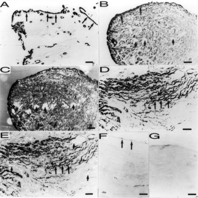
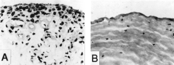
Methods: Alkaline phosphatase-anti-alkaline phosphatase (APAAP) method was used for immunohistochemical staining of tenascin-C in interface tissue and control synovial tissue.
Results: Tenascin-C was found to be a major component of the extracellular matrix at a hitherto unrecognised site, namely the synovial membrane-like interface tissue between implant and bone in aseptic loosening of THR. The overall tenascin-C staining (median score 4.0) was greatly increased in aseptic loosening compared with synovial membrane (median score 2.0; p < 0.001) and fibrous capsule (median score 2.0; p < 0.001) from primary THR operations. Topological analysis disclosed that tenascin-C was also found at the critical implant-interface and interface-bone surfaces.
Conclusion: Local tenascin-C expression is increased as a result of a chronic foreign body type reaction associated with excessive cytokine production and tissue injury mediated by microtrauma and neutral endoproteinases. This qualitative and topological deterioration of the bonding interlayer by an increase of anti-adhesive tenascin-C expression may inadvertantly contribute to loosening.
Figures
Similar articles
-
Production of platelet-derived growth factor in aseptic loosening of total hip replacement.Rheumatol Int. 1998;17(6):215-21. doi: 10.1007/s002960050037. Rheumatol Int. 1998. PMID: 9592860
-
Interleukin-11 (IL-11) in aseptic loosening of total hip replacement (THR).Scand J Rheumatol. 1998;27(5):363-7. doi: 10.1080/03009749850154393. Scand J Rheumatol. 1998. PMID: 9808400
-
Macrophage-colony stimulating factor (M-CSF) is increased in the synovial-like membrane of the periprosthetic tissues in the aseptic loosening of total hip replacement (THR).Clin Rheumatol. 1997 May;16(3):243-8. doi: 10.1007/BF02238958. Clin Rheumatol. 1997. PMID: 9184260 Clinical Trial.
-
[Wear particles: key to aseptic prosthetic loosening?].Pathologe. 2006 Nov;27(6):447-60. doi: 10.1007/s00292-006-0868-4. Pathologe. 2006. PMID: 17024444 Review. German.
-
[Particle disease--aseptic loosening of the total hip endoprosthesis].Lijec Vjesn. 2008 Jan-Feb;130(1-2):16-20. Lijec Vjesn. 2008. PMID: 18589638 Review. Croatian.
Cited by
-
The calreticulin-binding sequence of thrombospondin 1 regulates collagen expression and organization during tissue remodeling.Am J Pathol. 2010 Oct;177(4):1710-24. doi: 10.2353/ajpath.2010.090903. Epub 2010 Aug 19. Am J Pathol. 2010. PMID: 20724603 Free PMC article.
References
Publication types
MeSH terms
Substances
LinkOut - more resources
Full Text Sources
Medical
