

Transplanted and repopulated retinal pigment epithelial cells on damaged Bruch's membrane in rabbits
- PMID: 9893598
- PMCID: PMC1722745
- DOI: 10.1136/bjo.82.9.1056
Transplanted and repopulated retinal pigment epithelial cells on damaged Bruch's membrane in rabbits
Abstract
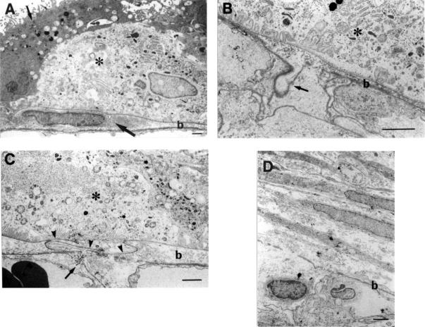
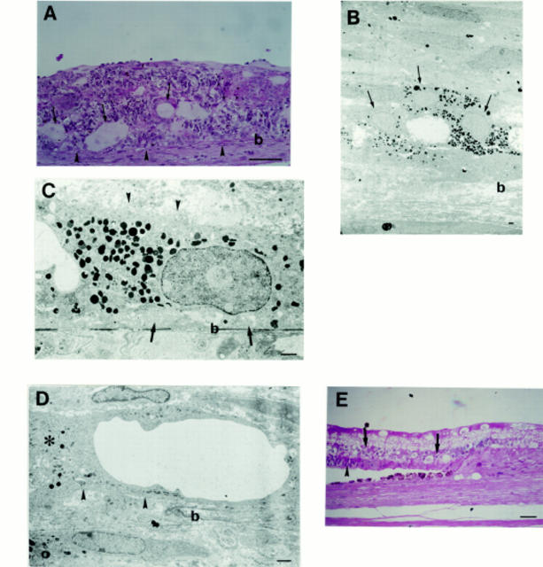
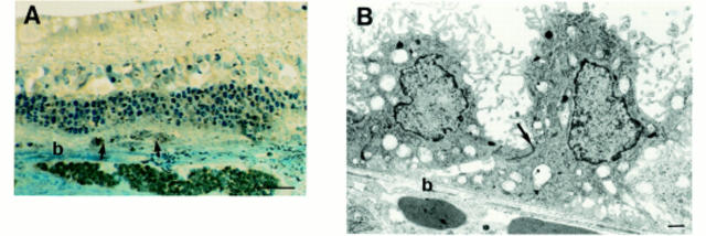
Aims: The authors studied how artificially damaged Bruch's membrane influenced growth and differentiation of transplanted embryonic retinal pigment epithelial (RPE) cells and of host RPE cells in rabbits.
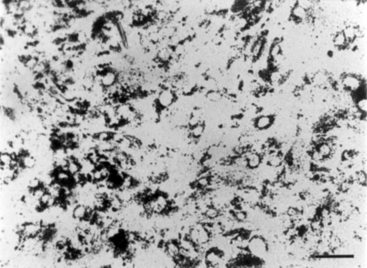
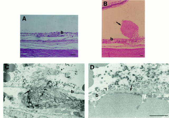
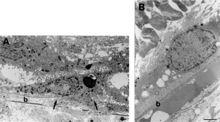
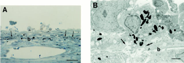
Methods: Embryonic RPE cells obtained from pigmented rabbits were transplanted into the subretinal space of adult albino rabbits. The host RPE was removed with a silicone cannula, and Bruch's membrane was damaged by scratching with a microhooked 27 gauge needle under the detached retina in closed vitrectomy. The transplantation sites were examined 3, 7, and 14 days after surgery by light and electron microscopy.
Results: Varying degrees of damage in Bruch's membrane were observed. Pigmented and hypopigmented RPE cells showed a normal polarity and tight junctions were seen at the sites of mild to moderate damage 3-7 days after the surgery. In contrast, fibroblast-like cells with no such features of RPE cells formed multiple layers at the sites of severe damage involving the full thickness of Bruch's membrane and the choriocapillaris even 14 days after the surgery. Without transplantation, host RPE cells repopulated the damaged areas in the same way as transplanted RPE cells.
Conclusions: Transplanted embryonic RPE cells as well as host RPE cells grew and differentiated on the moderately damaged Bruch's membrane, while the severely damaged Bruch's membrane did not allow differentiation of RPE cells although these cells could grow and cover the damaged areas.
Figures
Similar articles
-
In vitro transplantation of fetal human retinal pigment epithelial cells onto human cadaver Bruch's membrane.Exp Eye Res. 1998 Jan;66(1):49-67. doi: 10.1006/exer.1997.0404. Exp Eye Res. 1998. PMID: 9533831
-
Autologous transplantation of retinal pigment epithelium after mechanical debridement of Bruch's membrane.Curr Eye Res. 2003 Feb;26(2):81-8. doi: 10.1076/ceyr.26.2.81.14508. Curr Eye Res. 2003. PMID: 12815526
-
Impaired RPE survival on aged submacular human Bruch's membrane.Exp Eye Res. 2005 Feb;80(2):235-48. doi: 10.1016/j.exer.2004.09.006. Exp Eye Res. 2005. PMID: 15670802
-
Improving RPE adhesion to Bruch's membrane.Eye (Lond). 2009 Oct;23(10):1890-3. doi: 10.1038/eye.2008.411. Epub 2009 Jan 16. Eye (Lond). 2009. PMID: 19151642 Review.
-
Biochemical restoration of aged human Bruch's membrane: experimental studies to improve retinal pigment epithelium transplant survival and differentiation.Dev Ophthalmol. 2014;53:133-42. doi: 10.1159/000358531. Epub 2014 Apr 10. Dev Ophthalmol. 2014. PMID: 24732767 Review.
Cited by
-
Age-related macular degeneration: epidemiology and optimal treatment.Drugs Aging. 2002;19(2):101-33. doi: 10.2165/00002512-200219020-00003. Drugs Aging. 2002. PMID: 11950377 Review.
-
Localized Structural and Functional Deficits in a Nonhuman Primate Model of Outer Retinal Atrophy.Invest Ophthalmol Vis Sci. 2021 Oct 4;62(13):8. doi: 10.1167/iovs.62.13.8. Invest Ophthalmol Vis Sci. 2021. PMID: 34643661 Free PMC article.
-
Porous poly(ε-caprolactone) scaffolds for retinal pigment epithelium transplantation.Invest Ophthalmol Vis Sci. 2014 Mar 25;55(3):1754-62. doi: 10.1167/iovs.13-12833. Invest Ophthalmol Vis Sci. 2014. PMID: 24550370 Free PMC article.
-
A novel rabbit model for studying RPE transplantation.Invest Ophthalmol Vis Sci. 2008 Sep;49(9):4115-25. doi: 10.1167/iovs.08-1976. Epub 2008 May 23. Invest Ophthalmol Vis Sci. 2008. PMID: 18502985 Free PMC article.
-
Basement membranes and artificial substrates in cell transplantation.Graefes Arch Clin Exp Ophthalmol. 2004 Jan;242(1):68-75. doi: 10.1007/s00417-003-0800-z. Epub 2003 Nov 20. Graefes Arch Clin Exp Ophthalmol. 2004. PMID: 14628146 Review. No abstract available.
References
Publication types
MeSH terms
LinkOut - more resources
Full Text Sources