. 1998 Sep;82(9):1063-71.
doi: 10.1136/bjo.82.9.1063.

Expression of cell adhesion molecules and vascular endothelial growth factor in experimental choroidal neovascularisation in the rat

Affiliations

Expression of cell adhesion molecules and vascular endothelial growth factor in experimental choroidal neovascularisation in the rat

W Y Shen et al. Br J Ophthalmol. 1998 Sep.

Abstract

Aims: To investigate the longevity and reproducibility of choroidal neovascularisation (CNV) induced by krypton laser photocoagulation in the rat. The presence of cell adhesion molecules (CAMs) and vascular endothelial growth factor (VEGF) during the development of CNV was also studied.

Methods: 67 pigmented rats underwent retinal photocoagulation by krypton laser. The eyes were examined by either single or serial fluorescein angiography at 3 days, 1, 2-3, 4-5, 7-8, and 12 weeks post photocoagulation. The expression of CAMs (ICAM-1, E-selectin, and CD44) and VEGF post photocoagulation was studied by immunohistochemistry.

Results: CNV related fluorescein leakage appeared in 46.4% of 766 laser spots delivered to the 58 eyes that were tested at 2-3 weeks post treatment. The ratio of hyperfluorescent laser sites did not change significantly at 8 weeks post laser. The number of leaky spots was independent of the total number of lesions delivered to each eye (at 2-3 weeks post laser 10-15 spots/eye: 44% and 25-30 spots/eye: 49%; t = 0.7673; p = 0.3903). Nine eyes were followed by serial angiography between 2 and 12 weeks. The laser spots with fluorescein leakage at 2 weeks (51.5%) remained leaky at 12 weeks (51.5%). Histopathologically, macrophage accumulation peaked at 5 days and CNV was firstly observed at 1 week post photocoagulation. ICAM-1, E-selectin, CD44, and VEGF were maximally induced at 3-5 days post laser photocoagulation, and were localised to RPE, choroidal vascular endothelial, and inflammatory cells. VEGF was also detected in intravascular leucocytes at the sites of laser lesions.

Conclusions: These studies demonstrated that krypton laser photocoagulation can be successfully used to produce lesions similar to those of human CNV. The response induced remained present for an extended period of time (12 weeks), thus offering a potential model to screen candidate CNV inhibitory agents. In addition, it is proposed that the expression of ICAM-1, E-selectin, CD44, and VEGF before new vessel formation might be linked to the initiation of CNV.


Figures

Figure 1
Figure 1
Choroidal neovascularisation induced by krypton laser photocoagulation in sample 3 of Table 2. (A) Fundus photograph demonstrating the 15 laser spots delivered. Laser spots were photographed at 3 days post photocoagulation and were consecutively numbered. (B-D) Fluorescein angiograms of the same animal taken at different time points after photocoagulation. (B) At 3 days post photocoagulation no fluorescein leakage was seen; however, some non-staining window defects were noted (arrows). (C) At 2 weeks post photocoagulation fluorescein leakage was detected in seven of the 15 laser spots. (D) At 3 months post photocoagulation the same laser spots maintained fluorescein leakage.
Figure 2
Figure 2
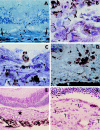
Histopathological studies after laser photocoagulation. (A) One week after photocoagulation, new vessels (small arrows) occurred with the disruption of Bruch's membrane (large arrows) (×400). (B) Three days post photocoagulation, RPE cells (open arrows) showed migration, proliferation, and multilayering. The infiltrating cells predominantly consisted of neutrophils (arrows) and small number of monocyte macrophages (m, arrowhead) (×1000). (C) Three months post photocoagulation, the neovascular network (arrows) disrupted the sealed RPE layer and invaded into the neural retina. Pigment laden macrophages (m) were observed around and within the region of neovascularisation and one macrophage (star) was located at the advancing front of the neovascular network (×1000). (D) Six weeks post photocoagulation, numerous CD68 positive macrophages (arrows) were observed in the laser photocoagulated area (×400). (E) Three months after photocoagulation, a section from the eye of Figure 1D demonstrates a fully developed neovascular membrane (star) (×100). (F) Higher magnification of (E) demonstrated numerous new vessels (arrows) within the neovascular membrane (×400). (A) and (C) Resin embedded, stained with toluidine blue. (B), (E), and (F) Paraffin sections stained with haematoxylin and eosin. (D) Cryostat section counterstained with haematoxylin.
Figure 3
Figure 3
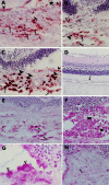
Immunohistochemical studies after laser photocoagulation. (A) Immunostaining for ICAM-1 at 3 days after photocoagulation (×400). (B) Immunostaining for E-selectin, 5 days after photocoagulation (×400). (C) Immunostaining for CD44, 5 days after photocoagulation (×400). In A-C, positive staining was localised to choroidal vascular endothelial cells (arrows), infiltrating cells (arrowheads), and RPE cells (open arrows). The star in (A) indicates the photocoagulated site. (D) The normal rat retina demonstrated very weak immunoreactivity to VEGF in RPE (arrow) and choroidal layer (×200). (E-H) Immunostaining for VEGF at 5 days after photocoagulation (×400). Following photocoagulation, positive staining was localised to the choroidal vascular endothelial cells (E, arrows), accumulating infiltrating cells (F, arrowheads), and the proliferating RPE cells (G, open arrows). No VEGF immunostaining was detected in the foci of lesion where neovascularisation has been enveloped by the proliferating cells (H). Arrow is pointing to a new vessel in the middle of a neovascular membrane.

References

    1. Am J Ophthalmol. 1992 Oct 15;114(4):464-72 - PubMed
    1. Eur J Ophthalmol. 1992 Apr-Jun;2(2):51-7 - PubMed
    1. J Exp Med. 1990 Dec 1;172(6):1535-45 - PubMed
    1. Invest Ophthalmol Vis Sci. 1986 Nov;27(11):1644-52 - PubMed
    1. Graefes Arch Clin Exp Ophthalmol. 1987;225(1):70-6 - PubMed

Publication types

MeSH terms