Comparative Study
. 1998 Dec;107(6-7):366-75.
doi: 10.1007/s004120050320.

A new look at kinetochore structure in vertebrate somatic cells using high-pressure freezing and freeze substitution

Affiliations
Comparative Study

A new look at kinetochore structure in vertebrate somatic cells using high-pressure freezing and freeze substitution

B F McEwen et al. Chromosoma. 1998 Dec.

Abstract

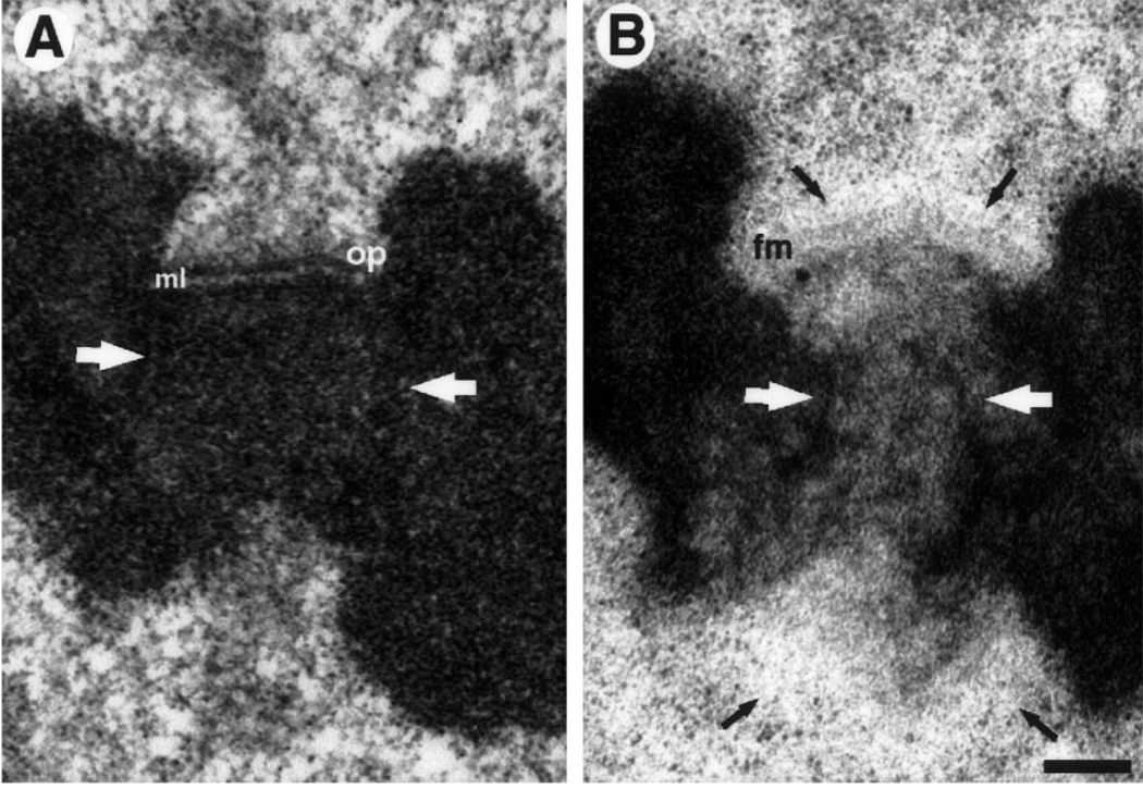
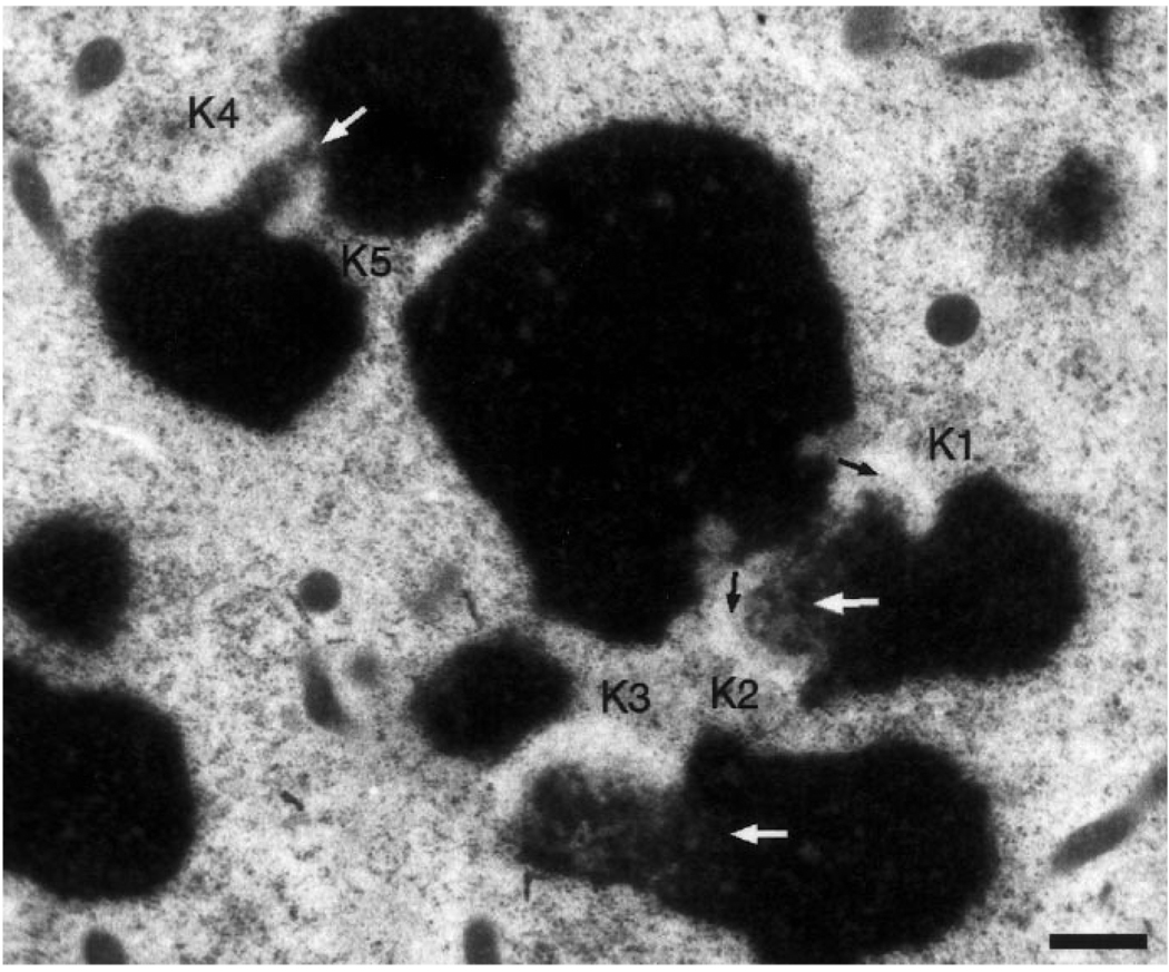
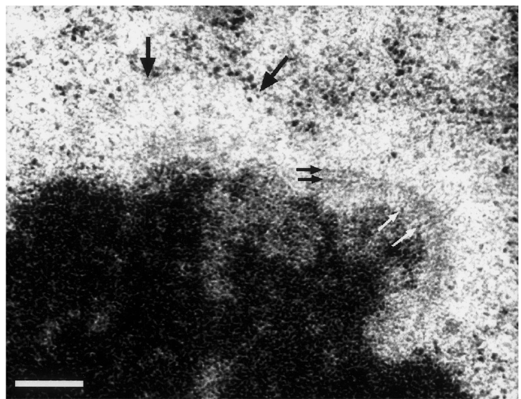
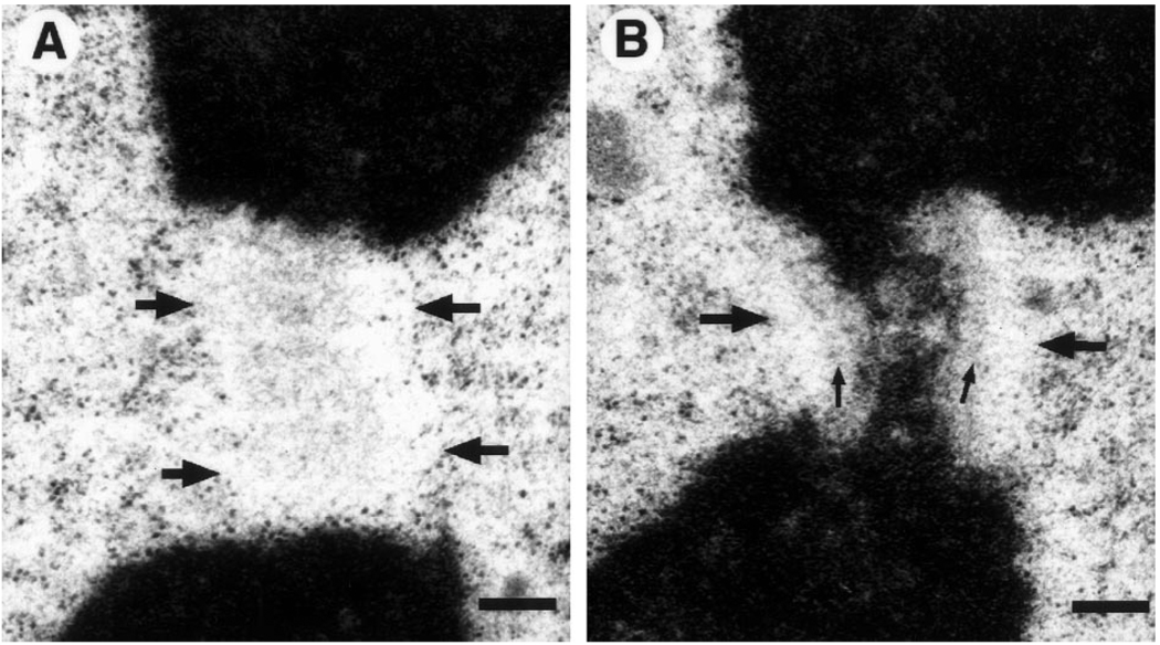
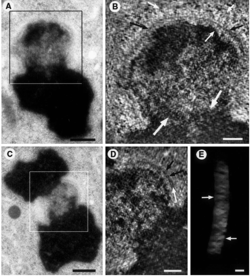
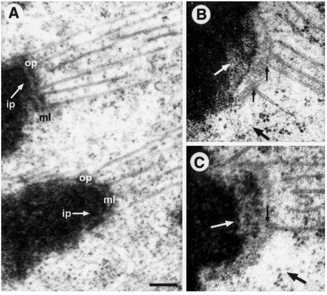
Three decades of structural analysis have produced the view that the kinetochore in vertebrate cells is a disk-shaped structure composed of three distinct structural domains. The most prominent of these consists of a conspicuous electron opaque outer plate that is separated by a light-staining electron-translucent middle plate from an inner plate associated with the surface of the pericentric heterochromatin. Spindle microtubules terminate in the outer plate and, in their absence, a conspicuous corona of fine filaments radiates from the cytoplasmic surface of this plate. Here we report for the first time the ultrastructure of kinetochores in untreated and Colcemid-treated vertebrate somatic (PtK1) cells prepared for optimal structural preservation using high-pressure freezing and freeze substitution. In serial thin sections, and electron tomographic reconstructions, the kinetochore appears as a 50-75 nm thick mat of light-staining fibrous material that is directly connected with the more electron-opaque surface of the centromeric heterochromatin. This mat corresponds to the outer plate in conventional preparations, and is surrounded on its cytoplasmic surface by a conspicuous 100-150 nm wide zone that excludes ribosomes and other cytoplasmic components. High magnification views of this zone reveal that it contains a loose network of light-staining, thin (<9 nm diameter) fibers that are analogous to the corona fibers in conventional preparations. Unlike the chromosome arms, which appear uniformly electron opaque, the chromatin in the primary constriction appears mottled. Since the middle plate is not visible in these kinetochore preparations this feature is likely an artifact produced by extraction and coagulation during conventional fixation and/or dehydration procedures.


Figures

Fig. 1
Fig. 1
A, B. Comparison between high-pressure freezing/freeze substitution (HPF/FS) and conventional specimen preparations for Colcemid treated PtK1 cells. A Kinetochore prepared via conventional protocol. The outer plate (op) is a heavily stained, compact, structure that is separated from the underlying heterochromatin by a translucent middle layer (ml). A prominent fibrous corona radiates from the outer plate’s distal surface. Note the general extracted appearance of the surrounding cytoplasm, and the uniform staining of the centromeric chromatin (white arrows). B Sister kinetochores prepared via HPF/FS. In the top kinetochore, the fibrous mat structure (fm) is lightly stained and much more open than the outer plate in A. The corona appears as a cytoplasmic exclusion zone (black arrows) lacking in discernable substructure. In contrast to conventional preparations, the heterochromatin has a mottled appearance (white arrows) and the surrounding cytoplasm is smooth, uniform, and unextracted. The lower kinetochore is sectioned at an oblique angle. As a result the mat structure is not evident, and was not found in the neighboring serial sections. However, the exclusion zone is visible (arrows). Bar represents 250 nm
Fig. 2
Fig. 2
Low magnification view of a Colcemid-treated PtK1 cell prepared via HPF/FS. Kinetochores are most readily recognized by the adjoining clear zones that exclude ribosomes and other cytoplasmic particles. Longitudinally sectioned kinetochores (K1, K2) also show faintly staining mats (black arrows) that are not evident in obliquely sectioned kinetochores (K3-K5). The mottled appearance of centromeric heterochromatin (white arrows) is a general feature of chromosomes prepared via HPF/FS. Bar represents 500 nm
Fig. 3
Fig. 3
Higher magnification view of a Colcemid-treated HPF/FS kinetochore. This chromosome is oriented with a slight rotation into the section plane. The mat structure on the right-hand kinetochore appears to be constructed from fine fibers radiating out from the underlying chromatin (white arrows) and crossing fibers lying parallel to the chromatin surface (black arrows). The mat is not visible in the left-hand kinetochore in this section, but the kinetochore can be detected by the prominent exclusion zone with ribosomes piled up along the boundary (large black arrows). Bar represents 200 nm
Fig. 4
Fig. 4
A, B. The exclusion zone in an obliquely cut section. A A roughly axial view of the exclusion zone (boundaries marked by large arrows), detected primarily by exclusion of ribosomes and other cytoplasmic material. A fine flocculent material, possibly of fibrous nature, is seen in this area. Presumably this material consists of corona fibers and/or an en face view of the mat structure B Serial section to A, showing the obliquely sectioned kinetochore. Both the mat structure (small arrows) and exclusion zone (large arrows) can be discerned. Bar represents 250 nm
Fig. 5
Fig. 5
A–E. Tomographic reconstructions of colcemid-treated, HPF/FS kinetochores. A Low magnification view of sister kinetochore used for tomographic reconstruction (box indicates the approximate area of the reconstruction shown in B). As in Fig. 3, the chromosome is oriented with a slight rotation into the section plane. Bar represents 500 nm. B A 9 nm thick slice (average of three successive 2.9 nm thick tomograms) from the three-dimensional reconstruction of the boxed area in A. Fibrous elements are evident radiating from the heterochromatin and from the surface of the mat (small white arrows) but low contrast and inherent resolution limits preclude exact measurements of diameters and lengths. In this view the mat structure appears as a long, thin, fiber-like element running parallel to the chromatin surface (black arrows). A striking difference in texture between euchromatin and centromeric heterochromatin is evident along the border of the two domains (large white arrows). Bar represents 200 nm. C Low magnification view of the kinetochore used for the tomographic reconstruction illustrated in D and E (box delineates the reconstructed area). Bar represents 500 nm. D A 9 nm thick slice (like B) from the three-dimensional reconstruction of the boxed area in D. The mat shows a block-like arrangement (black arrows) and some unit blocks having a tripartite organization (white arrows). Bar represents 200 nm. E Tilted view of the mat structure. The mat has been isolated from the remainder of the three-dimensional reconstruction and is displayed as a semi-transparent subvolume using masking tools and volume rendering software (see McEwen et al. 1993; McEwen and Marko 1998). The block-like arrangement of putative unit structures is particularly evident in this view (arrows). Bar represents 50 nm
Fig. 6
Fig. 6
A–C. HPF/FS vs conventional specimen preparations for untreated PtK1 cells. A Adjacent kinetochores from a conventionally prepared metaphase cell. Note the prominent outer plate (op) structure that stains as heavily as chromatin, and is separated from the underlying inner plate (ip) by a well-defined, translucent, middle layer (ml).Microtubule (Mt) plus ends can not be clearly traced within the highly condensed outer plate. B, C Two different kinetochores from an anaphase cell prepared via HPF/FS. Note the absence of a prominent outer plate. Instead there is a lightly staining mat structure that sits above the chromatin without an intervening translucent layer. Mt plus ends can be traced (small black arrows) and they appear to flay apart and be continuous with the fibrous mat (in agreement with Mastronarde et al. 1997). The cytoplasmic exclusion zone can still be detected in some areas where there are no Mts (large black arrows). The mottled appearance of the centromeric heterochromatin is also evident (white arrows). Bar represents 200 nm

References

    1. Bernat RL, Delannoy MR, Rothfield NF, Earnshaw WC. Disruption of centromere assembly during interphase inhibits kinetochore morphogenesis and function in mitosis. Cell. 1991;66:1229–1238. - PubMed
    1. Brinkley BR, Stubblefield E. Fine structure of the kinetochore in Chinese hamster cells in vitro. Chromosoma. 1966;19:28–43. - PubMed
    1. Campbell MS, Gorbsky GJ. Microinjection of mitotic cells with the 3F3/2 antiphosphoepitope antibody delays the onset of anaphase. J Cell Biol. 1995;129:1195–1204. - PMC - PubMed
    1. Cassimeris L, Rieder CL, Rupp G, Salmon ED. Stability of microtubule attachment to metaphase kinetochores in PtK1 cells. J Cell Sci. 1990;96:9–15. - PubMed
    1. Chen R-H, Waters JC, Salmon ED, Murray AW. Association of spindle assembly checkpoint component xMAD2 with unattached kinetochores. Science. 1996;274:242–246. - PubMed

Publication types

Substances

LinkOut - more resources