

An in vitro tissue culture bilayer model to examine early events in Mycobacterium tuberculosis infection
- PMID: 9916072
- PMCID: PMC96368
- DOI: 10.1128/IAI.67.2.653-658.1999
An in vitro tissue culture bilayer model to examine early events in Mycobacterium tuberculosis infection
Abstract
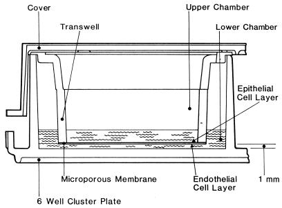
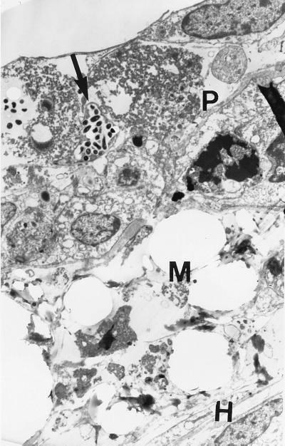
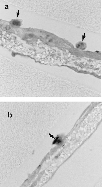
A tissue culture bilayer system that mimics some aspects of early alveolar infection by Mycobacterium tuberculosis was developed. This model incorporates human lung epithelial type II pneumocyte (A549) (upper chamber) and endothelial cell (lower chamber) layers separated by a microporous membrane. This construction makes it possible to observe and quantify the passage of bacteria through the two layers, to observe the interaction of the bacteria with the various cell types, and to examine the basic mechanisms of immune cell recruitment to the site of infection. After 10(7) organisms were added to the upper chamber we microscopically observed large numbers of bacteria attached to and within the pneumocytes and we determined by viable-cell counting that a small percentage of the inoculum (0.02 to 0.43%) passed through the bilayer into the lower chamber. When peripheral blood mononuclear cells were added to the lower chamber, microscopic examination indicated a migration of the mononuclear cells through the bilayer to the apical surface, where they were seen associated with the mycobacteria on the pneumocytes. The added complexity of the bilayer system offers an opportunity to define more precisely the roles of the various lung cell types in the pathogenesis of early tuberculosis.
Figures

References
-
- Bloom B R, Fine P E M. The BCG experience: implications for future vaccines against tuberculosis. In: Bloom B R, editor. Tuberculosis: pathogenesis, protection, and control. Washington, D.C: ASM Press; 1994. pp. 531–558.
-
- Burkitt H G, Young B, Heath J W. Wheater’s functional histology: a text and colour atlas. Edinburgh, Scotland: Churchill Livingstone; 1993.
-
- Castro-Garza, J. C., et. al. Unpublished data.
MeSH terms
LinkOut - more resources
Full Text Sources
Other Literature Sources
