

Characterization of the avian pathogenic Escherichia coli hemagglutinin Tsh, a member of the immunoglobulin A protease-type family of autotransporters
- PMID: 9916089
- PMCID: PMC96385
- DOI: 10.1128/IAI.67.2.772-781.1999
Characterization of the avian pathogenic Escherichia coli hemagglutinin Tsh, a member of the immunoglobulin A protease-type family of autotransporters
Abstract
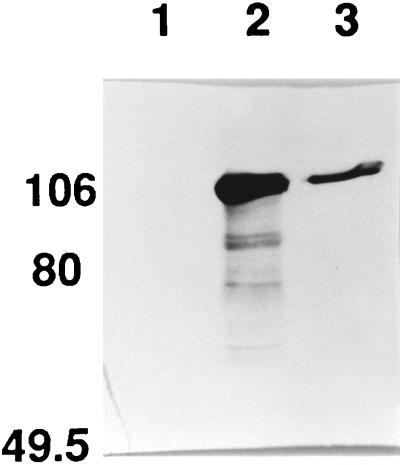
We reported earlier that a single gene, tsh, isolated from a strain of avian pathogenic Escherichia coli (APEC) was sufficient to confer on E. coli K-12 a hemagglutinin-positive phenotype and that the deduced sequence of the Tsh protein shared homology to the serine-type immunoglobulin A (IgA) proteases of Neisseria gonorrhoeae and Haemophilus influenzae. In this report we show that E. coli K-12 containing the recombinant tsh gene produced two proteins, a 106-kDa extracellular protein and a 33-kDa outer membrane protein, and was also able to agglutinate chicken erythrocytes. N-terminal sequence data indicated that the 106-kDa protein, designated Tshs, was derived from the N-terminal end of Tsh after the removal of a 52-amino-acid N-terminal signal peptide, while the 33-kDa protein, designated Tshbeta, was derived from the C-terminal end of Tsh starting at residue N1101. The Tshs domain contains the 7-amino-acid serine protease motif that includes the active-site serine (S259), found also in the secreted domains of the IgA proteases. However, site-directed mutagenesis of S259 did not abolish the hemagglutinin activity or the extracellular secretion of Tshs indicating that host-directed proteolysis was mediating the release of Tshs. Studies with an E. coli K-12 ompT mutant strain showed that the surface protease OmpT was not needed for the secretion of Tshs. Tsh belongs to a subclass of the IgA protease family, which also includes EspC of enteropathogenic E. coli, EspP of enterohemorragic E. coli, and SepA and VirG of Shigella flexneri, which seem to involve a host endopeptidase to achieve extracellular release of their N-terminal domains. In proteolytic studies conducted in vitro, Tshs did not cleave the substrate of the IgA proteases, human IgA1 or chicken IgA, and did not show proteolytic activity in a casein-based assay. Correlation of Tsh expression and hemagglutination activity appears to be a very complex phenomenon, influenced by strain and environmental conditions. Nevertheless, for both APEC and recombinant E. coli K-12 strains containing the tsh gene, it was only the whole bacterial cells and not the cell-free supernatants that could confer hemagglutinin activity. Our results provide insights into the expression, secretion, and proteolytic features of the Tsh protein, which belongs to the growing family of gram-negative bacterial extracellular virulence factors, named autotransporters, which utilize a self-mediated mechanism to achieve export across the bacterial cell envelope.
Figures


Similar articles
-
Functional analysis of the Tsh autotransporter from an avian pathogenic Escherichia coli strain.Infect Immun. 2004 Oct;72(10):5548-54. doi: 10.1128/IAI.72.10.5548-5554.2004. Infect Immun. 2004. PMID: 15385451 Free PMC article.
-
Isolation and characterization of a gene involved in hemagglutination by an avian pathogenic Escherichia coli strain.Infect Immun. 1994 Apr;62(4):1369-80. doi: 10.1128/iai.62.4.1369-1380.1994. Infect Immun. 1994. PMID: 8132344 Free PMC article.
-
Characterization of EspC, a 110-kilodalton protein secreted by enteropathogenic Escherichia coli which is homologous to members of the immunoglobulin A protease-like family of secreted proteins.J Bacteriol. 1996 Nov;178(22):6546-54. doi: 10.1128/jb.178.22.6546-6554.1996. J Bacteriol. 1996. PMID: 8932311 Free PMC article.
-
Serine protease autotransporters of enterobacteriaceae (SPATEs): biogenesis and function.Toxins (Basel). 2010 Jun;2(6):1179-206. doi: 10.3390/toxins2061179. Epub 2010 May 28. Toxins (Basel). 2010. PMID: 22069633 Free PMC article. Review.
-
The secretion pathway of IgA protease-type proteins in gram-negative bacteria.Bioessays. 1993 Dec;15(12):799-805. doi: 10.1002/bies.950151205. Bioessays. 1993. PMID: 8141798 Review.
Cited by
-
From self sufficiency to dependence: mechanisms and factors important for autotransporter biogenesis.Nat Rev Microbiol. 2012 Feb 16;10(3):213-25. doi: 10.1038/nrmicro2733. Nat Rev Microbiol. 2012. PMID: 22337167 Review.
-
AatA is a novel autotransporter and virulence factor of avian pathogenic Escherichia coli.Infect Immun. 2010 Mar;78(3):898-906. doi: 10.1128/IAI.00513-09. Epub 2009 Dec 22. Infect Immun. 2010. PMID: 20028805 Free PMC article.
-
Three new serine-protease autotransporters of Enterobacteriaceae (SPATEs) from extra-intestinal pathogenic Escherichia coli and combined role of SPATEs for cytotoxicity and colonization of the mouse kidney.Virulence. 2019 Dec;10(1):568-587. doi: 10.1080/21505594.2019.1624102. Virulence. 2019. PMID: 31198092 Free PMC article.
-
Transfer region of pO113 from enterohemorrhagic Escherichia coli: similarity with R64 and identification of a novel plasmid-encoded autotransporter, EpeA.Infect Immun. 2003 Nov;71(11):6307-19. doi: 10.1128/IAI.71.11.6307-6319.2003. Infect Immun. 2003. PMID: 14573650 Free PMC article.
-
Intramolecular interactions between the protease and structural domains are important for the functions of serine protease autotransporters.Infect Immun. 2010 Aug;78(8):3335-45. doi: 10.1128/IAI.00129-10. Epub 2010 May 17. Infect Immun. 2010. PMID: 20479079 Free PMC article.
References
-
- Bachovin W W, Plaut A G, Flentke G R, Lynch M, Kettner C A. Inhibition of IgA1 proteinases from Neisseria gonorrhoeae and Hemophilus influenzae by peptide prolyl boronic acids. J Biol Chem. 1990;265:3738–3743. - PubMed
-
- Bailey J M, Miller C G. Current protocols in protein science, unit 15.1. New York, N.Y: Wiley; 1996. C-terminal sequence analysis.
-
- Benjelloun-Touimi Z, Sansonetti P J, Parsot C. SepA, the major extracellular protein of Shigella flexneri: autonomous secretion and involvement in tissue invasion. Mol Microbiol. 1995;17:123–135. - PubMed
Publication types
MeSH terms
Substances
LinkOut - more resources
Full Text Sources
Miscellaneous
