. 1999 Feb;67(2):853-61.
doi: 10.1128/IAI.67.2.853-861.1999.

Expression and distribution of leptospiral outer membrane components during renal infection of hamsters

Affiliations

Expression and distribution of leptospiral outer membrane components during renal infection of hamsters

J K Barnett et al. Infect Immun. 1999 Feb.

Abstract

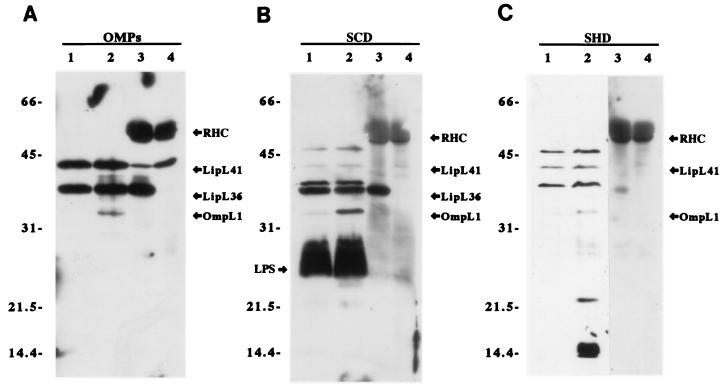
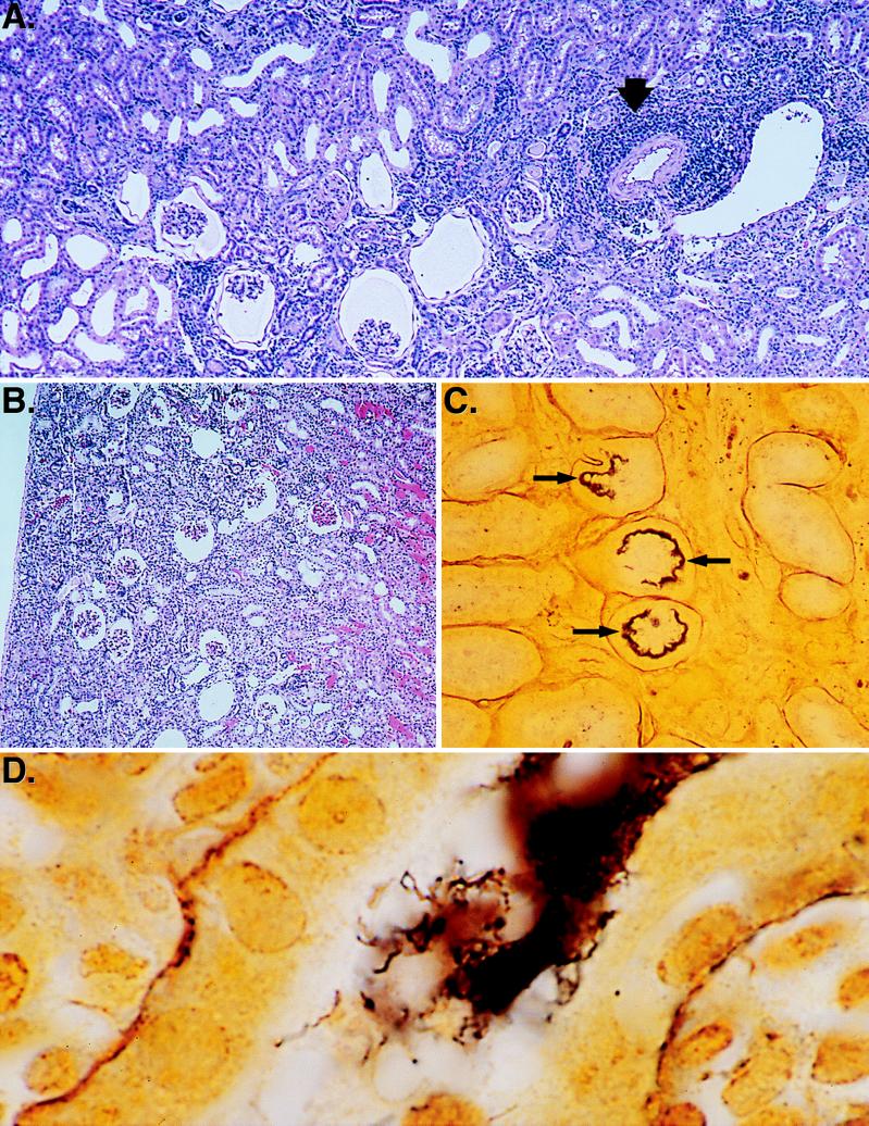
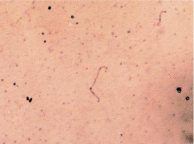
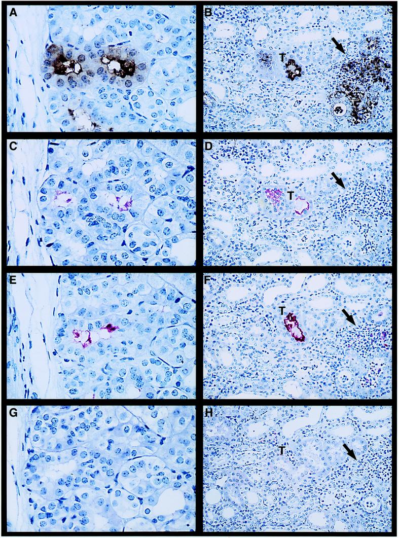
The outer membrane of pathogenic Leptospira species grown in culture media contains lipopolysaccharide (LPS), a porin (OmpL1), and several lipoproteins, including LipL36 and LipL41. The purpose of this study was to characterize the expression and distribution of these outer membrane antigens during renal infection. Hamsters were challenged with host-derived Leptospira kirschneri to generate sera which contained antibodies to antigens expressed in vivo. Immunoblotting performed with sera from animals challenged with these host-derived organisms demonstrated reactivity with OmpL1, LipL41, and several other proteins but not with LipL36. Although LipL36 is a prominent outer membrane antigen of cultivated L. kirschneri, its expression also could not be detected in infected hamster kidney tissue by immunohistochemistry, indicating that expression of this protein is down-regulated in vivo. In contrast, LPS, OmpL1, and LipL41 were demonstrated on organisms colonizing the lumen of proximal convoluted renal tubules at both 10 and 28 days postinfection. Tubular epithelial cells around the luminal colonies had fine granular cytoplasmic LPS. When the cellular inflammatory response was present in the renal interstitium at 28 days postinfection, LPS and OmpL1 were also detectable within interstitial phagocytes. These data establish that outer membrane components expressed during infection have roles in the induction and persistence of leptospiral interstitial nephritis.


Figures

FIG. 1
FIG. 1
Immunoblots of leptospiral proteins using antisera from hamsters challenged with culture-derived and host-derived L. kirschneri. Each panel is an immunoblot of leptospiral proteins that were unheated (lanes 1), boiled (lanes 2), and immunoprecipitated with antisera specific for LipL36 (lanes 3) and LipL41 (lanes 4). Panels were probed with a mixture of OmpL1, LipL36, and LipL41 antisera (A), an SCD sample (B), and an SHD sample (C). The SCD and SHD immunoblots shown are the results for serum from one animal from each group but are representative of immunoblot results obtained with sera from the other animals in the SCD and SHD groups. Hamster serum from uninfected littermates was nonreactive (data not shown). Rabbit heavy-chain (RHC) and light-chain immunoglobulin bands are visible in lanes 3 and 4 because these samples are immunoprecipitates.
FIG. 2
FIG. 2
Histopathology of hamster kidney after infection with L. kirschneri. (A) H&E stain of hamster kidney tissue 28 days after infection showing contracted glomeruli and an interstitial inflammatory infiltrate (arrow). Film magnification, ×25. Final magnification, ×150. (B) H&E stain of hamster kidney tissue 10 days after infection showing contracted glomeruli, vascular congestion, and proteinaceous debris in the tubules. Film magnification, ×25. Final magnification, ×100. (C) Silver stain of hamster kidney tissue 28 days after infection showing a low-power view of renal tubules with (arrows) and without spirochetal involvement. Film magnification, ×100. Final magnification, ×600. (D) Silver stain of hamster kidney tissue 28 days after infection showing a high-power view of the dense accumulation of spirochetes in a renal tubule. Film magnification, ×1,000. Final magnification, ×6,000.
FIG. 3
FIG. 3
Immunohistochemistry of cultivated L. kirschneri. A representative positive smear of cultivated L. kirschneri stained with rabbit polyclonal antiserum specific for LipL36 is shown. Film magnification, ×1,250. Final magnification, ×5,750.
FIG. 4
FIG. 4
Immunohistochemistry of kidney tissue obtained at 10 days (A, C, E, and G) (film magnification, ×400; final magnification, ×940) and 28 days (B, D, F, and H) (film magnification, ×160; final magnification, ×375) postinfection with virulent L. kirschneri by using the LPS-specific monoclonal antibody F71C2 (A and B) and rabbit polyclonal antisera specific for LipL41 (C and D), OmpL1 (E and F), and LipL36 (G and H). Higher magnification in the day 10 panels was needed to show the details of antigen expression in the renal tubular lumen and the presence of LPS in the cytoplasm of the tubular epithelial cells (A). No antigen was detected in the renal interstitium on day 10. A lower magnification was needed to show the wider distribution of antigen at the day 28 time point. LPS and OmpL1 were detected both in tubules (T) and in the renal interstitum (arrow) at the sites of inflammatory infiltrate (B and F, respectively).

References

    1. Alexander A D, Lessel E F, Evans L B, Franck E, Green S S. Preservation of leptospiras by liquid-nitrogen refrigeration. Int J Syst Bacteriol. 1972;22:165–169.
    1. Alves V A, Gayotto L C, Yasuda P H, Wakamatsu A, Kanamura C T, De Brito T. Leptospiral antigens (L. interrogans serogroup icterohaemorrhagiae) in the kidney of experimentally infected guinea pigs and their relation to the pathogenesis of the renal injury. Exp Pathol. 1991;42:81–93. - PubMed
    1. Alves V A F, Vianna M R, Yasuda P H, DeBrito T. Detection of leptospiral antigen in the human liver and kidney using an immunoperoxidase staining procedure. J Pathol. 1987;151:125–131. - PubMed
    1. Arean V M. The pathologic anatomy and pathogenesis of fatal human leptospirosis (Weil’s disease) Am J Pathol. 1962;40:393–423. - PMC - PubMed
    1. Auran N E, Johnson R C, Ritzi D M. Isolation of the outer sheath of Leptospira and its immunogenic properties in hamsters. Infect Immun. 1972;5:968–975. - PMC - PubMed

Publication types

MeSH terms