. 1999 Feb;67(2):914-20.
doi: 10.1128/IAI.67.2.914-920.1999.

Regulation of ExoS production and secretion by Pseudomonas aeruginosa in response to tissue culture conditions

Affiliations

Regulation of ExoS production and secretion by Pseudomonas aeruginosa in response to tissue culture conditions

A J Vallis et al. Infect Immun. 1999 Feb.

Abstract

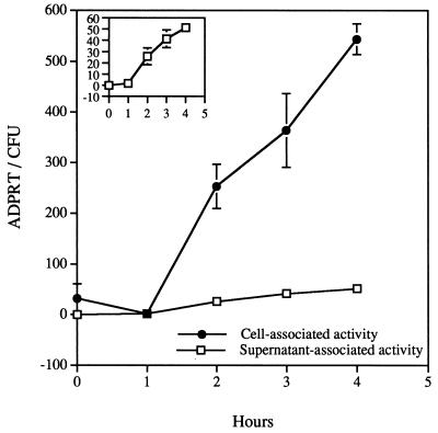
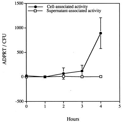
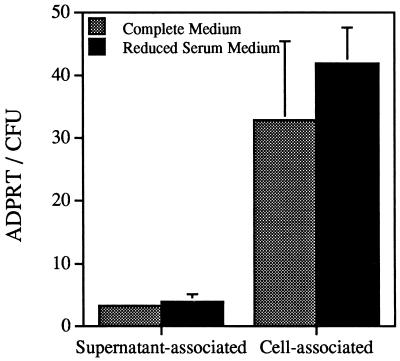
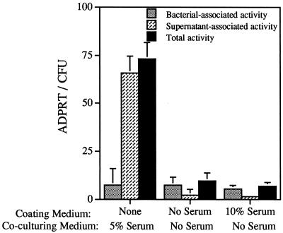
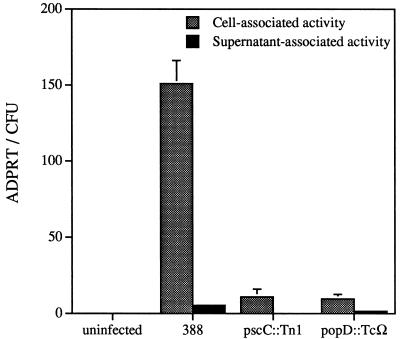
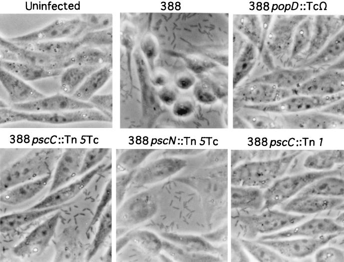
This study was initiated to characterize the regulation and secretion of ExoS by Pseudomonas aeruginosa during contact with eukaryotic cells. The production of ExoS was monitored by a sensitive ADP-ribosyltransferase activity assay, and specific activities were calculated for supernatant and cell-associated fractions. Time course analysis indicated that ExoS was produced after a lag period, suggesting that induction of the regulon is necessary for the expression of detectable amounts of enzyme activity. Under tissue culture growth conditions, ExoS was induced when P. aeruginosa was in contact with Chinese hamster ovary (CHO) cells or after growth in tissue culture medium with serum. The serum induction of ExoS appeared to result in generalized type III secretion, while induction by contact with CHO cells appeared to result in polarized type III secretion. Mutants in the type III secretory system that express a null phenotype for ExoS production in bacteriological medium produced but did not secrete the enzyme when P. aeruginosa was grown under inducing conditions in tissue culture medium. These results suggest that both induction and secretion of ExoS may differ when the bacteria are exposed to different growth environments. The putative type III translocation proteins and secretion apparatus of P. aeruginosa were required for translocation of bacterial factors that mediate changes in CHO cell morphology during infection.


Figures

FIG. 1
FIG. 1
Cell- and supernatant-associated ExoS ADP-ribosyltransferase activity from infections performed in the presence of serum. Cytochalasin D-treated CHO cells were infected with P. aeruginosa 388. Fractions consisting of soluble supernatant material (supernatant-associated activity) and CHO cell lysates (cell-associated activity) were collected and processed as detailed in Materials and Methods. ExoS activity is reported as 10−19 moles of ADP-ribosyltransferase activity that is normalized to CFUs and is presented as the mean and standard deviation of experiments performed in triplicate. The inset of Fig. 1 changes the scale of the y axis to demonstrate the accumulation of ExoS activity in supernatant fractions.
FIG. 2
FIG. 2
Cell- and supernatant-associated ExoS ADP-ribosyltransferase activity from infections performed in the absence of serum. Cytochalasin D-treated CHO cells were infected with P. aeruginosa 388. Fractions consisting of soluble supernatant material (supernatant-associated activity) and CHO cell lysates (cell-associated activity) were collected and processed as detailed in Materials and Methods. ExoS activity is reported as 10−19 moles of ADP-ribosyltransferase activity that is normalized to CFUs and is presented as the mean and standard deviation of experiments performed in triplicate. ExoS activity in supernatant-associated fractions was not detected.
FIG. 3
FIG. 3
Comparison of ExoS activity from CHO cells propagated in complete (10% serum) or reduced (0.15%) serum medium. CHO cells were cultured in complete medium with 10% serum (gray bar) or adapted by sequential passage in CHO III protein-free medium with progressive reduction in the amount of complete medium added. The final concentration of serum components that resulted in consistent CHO cell growth and viability was 0.15%. A bacterial inoculum was prepared in serum-free medium, and the cocultivation was allowed to proceed for 4 h. Fractions were prepared, and ExoS ADP-ribosyltransferase activity was normalized to CFUs as described in Materials and Methods.
FIG. 4
FIG. 4
The effect of adsorbed serum components on ExoS production. Tissue culture wells (in the absence of CHO cells) were left untreated or were pretreated overnight at 37°C with medium either containing or not containing newborn calf serum. The wells were washed once with PBS, and a bacterial inoculum with or without serum was added to each well. After a 4-h incubation period, the culture was harvested, and ExoS activity was measured in bacterial lysates (bacterium-associated activity) or in supernatant (supernatant-associated activity) fractions. The total activity measured is the sum of the bacterium- and supernatant-associated activities.
FIG. 5
FIG. 5
Cell- and supernatant-associated ExoS activities of P. aeruginosa strains. Wild-type (388) or mutant P. aeruginosa strains defective in type III secretion (pscC::Tn1) or translocation (popD::TcΩ) were cocultured with cytochalasin D-treated CHO cells for 4 h in serum-free medium. Cellular fractions were prepared, and ExoS activity was measured and normalized to CFUs as described in Materials and Methods.
FIG. 6
FIG. 6
Phase-contrast micrographs of CHO cells infected with parental strain 388 and isogenic mutant strains of P. aeruginosa (popD::TcΩ, pscC::Tn5Tc, pscN::Tn5Tc, and pscC::Tn1). CHO cells were left uninfected or were infected for 4 h with various bacterial strains in the absence of serum. Cells were washed, fixed, and photographed to assess cellular morphology in response to infection.

Similar articles

Cited by

References

    1. Aitken A, Jones D, Soneji Y, Howell S. 14-3-3 proteins: biological function and domain structure. Biochem Soc Trans. 1995;23:605–611. - PubMed
    1. Bjorn M J, Pavlovskis O R, Thompson M R, Iglewski B H. Production of exoenzyme S during Pseudomonas aeruginosa infections of burned mice. Infect Immun. 1979;24:837–842. - PMC - PubMed
    1. Boland A, Sory M P, Iriarte M, Kerbourch C, Wattiau P, Cornelis G R. Status of YopM and YopN in the Yersinia Yop virulon: YopM of Y. enterocolitica is internalized inside the cytosol of PU5-1.8 macrophages by the YopB,D,N delivery apparatus. EMBO J. 1996;15:5191–5201. - PMC - PubMed
    1. Coburn J. Pseudomonas aeruginosa exoenzyme S. Curr Top Microbiol Immunol. 1992;175:133–143. - PubMed
    1. Coburn J, Dillon S T, Iglewski B H, Gill D M. Exoenzyme S of Pseudomonas aeruginosa ADP-ribosylates the intermediate filament protein vimentin. Infect Immun. 1989;57:996–998. - PMC - PubMed

Publication types

LinkOut - more resources