

Fas-FasL interaction involved in pathogenesis of ocular toxoplasmosis in mice
- PMID: 9916110
- PMCID: PMC96406
- DOI: 10.1128/IAI.67.2.928-935.1999
Fas-FasL interaction involved in pathogenesis of ocular toxoplasmosis in mice
Abstract
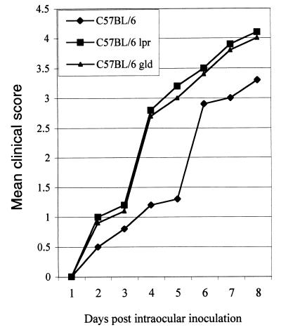
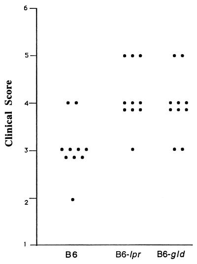
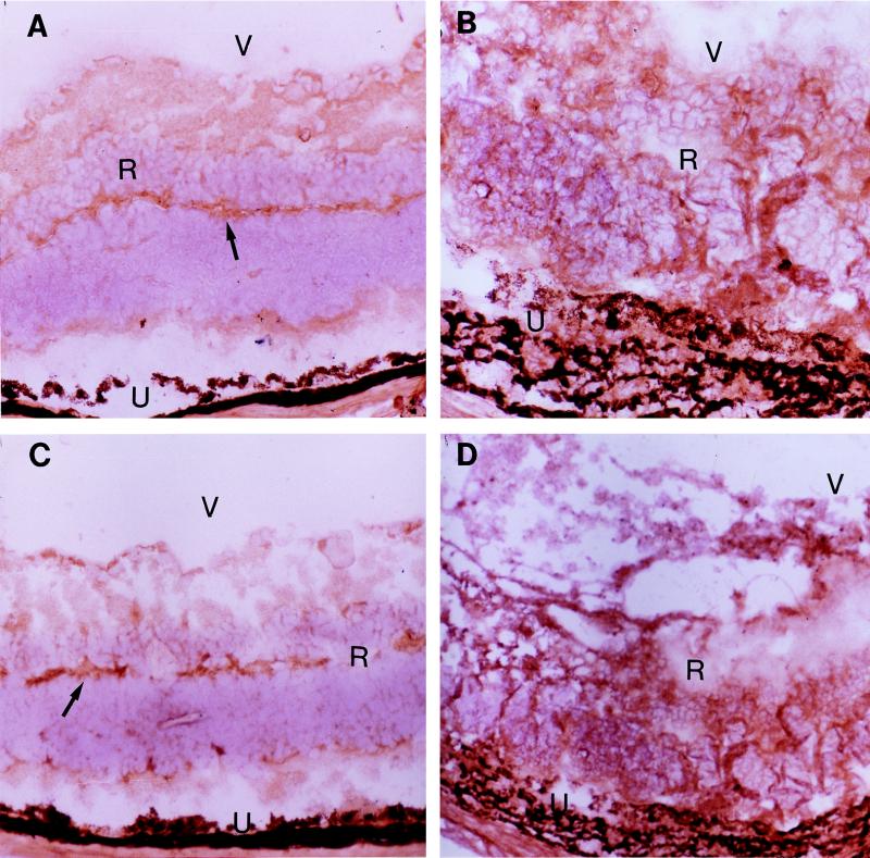
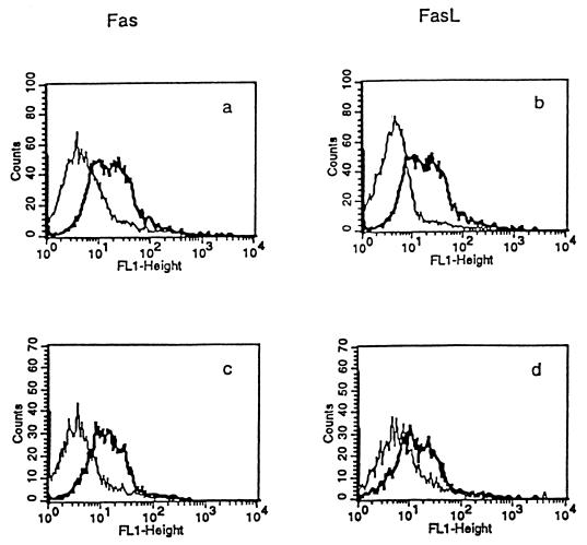
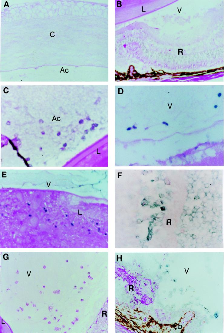
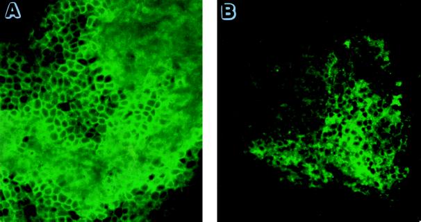
Ocular toxoplasmosis is a potentially blinding intraocular inflammation. The intent of this study was to investigate the role of Fas-FasL interaction in a murine model of acquired ocular toxoplasmosis induced by intracameral inoculation of Toxoplasma gondii. Intraocular inflammation, Fas and FasL expression on lymphocytes and on ocular tissues, the occurrence of apoptosis, and the frequency of CD8(+) and CD4(+) T cells in the infected eyes were analyzed in C57BL/6 (B6) mice. Susceptibility to parasite-induced intraocular inflammation was observed in Fas-deficient (B6-lpr) and FasL-deficient (B6-gld) mice. Inoculation of 5,000 T. gondii tachyzoites induced significant intraocular inflammation associated with increase of Fas and FasL expression in the inoculated eyes of wild-type B6 mice. Flow cytometry demonstrated a significant increase of Fas and FasL expression on the splenocytes from naive mice incubated in vitro with the parasite and on the splenocytes harvested from the infected mice at day 8 after parasite inoculation. Apoptosis of inflammatory cells and cells in ocular tissues was seen, and a greater frequency of CD8(+) than CD4(+) T cells was observed in the infected eyes. The intensity of intraocular inflammation was greater in B6-lpr and B6-gld mice than in wild-type B6 mice (P < 0.05). The results suggest that Fas-FasL interaction associated with apoptosis is involved in the pathogenesis of acquired ocular toxoplasmosis in mice.
Figures

Similar articles
-
Involvement of apoptosis and interferon-gamma in murine toxoplasmosis.Invest Ophthalmol Vis Sci. 2001 Aug;42(9):2031-6. Invest Ophthalmol Vis Sci. 2001. PMID: 11481268
-
Fas ligand (gld)- and Fas (lpr)-deficient mice do not show alterations in the extravasation or apoptosis of inflammatory neutrophils.J Leukoc Biol. 1998 Sep;64(3):373-83. doi: 10.1002/jlb.64.3.373. J Leukoc Biol. 1998. PMID: 9738665
-
Antiretroviral cytolytic T-lymphocyte nonresponsiveness: FasL/Fas-mediated inhibition of CD4(+) and CD8(+) antiviral T cells by viral antigen-positive veto cells.J Virol. 1999 May;73(5):3826-34. doi: 10.1128/JVI.73.5.3826-3834.1999. J Virol. 1999. PMID: 10196277 Free PMC article.
-
Analysis of gene profile, steady state proliferation and apoptosis of double-negative T cells in the periphery and gut epithelium provides new insights into the biological functions of the Fas pathway.Immunol Res. 2010 Jul;47(1-3):134-42. doi: 10.1007/s12026-009-8144-3. Immunol Res. 2010. PMID: 20066510 Free PMC article. Review.
-
Parasite dissemination and the pathogenesis of toxoplasmosis.Eur J Microbiol Immunol (Bp). 2011 Mar;1(1):3-9. doi: 10.1556/EuJMI.1.2011.1.3. Eur J Microbiol Immunol (Bp). 2011. PMID: 24466431 Free PMC article. Review. No abstract available.
Cited by
-
Toxoplasma gondii inhibits apoptosis in infected cells by caspase inactivation and NF-kappaB activation.Yonsei Med J. 2006 Dec 31;47(6):862-9. doi: 10.3349/ymj.2006.47.6.862. Yonsei Med J. 2006. PMID: 17191317 Free PMC article.
-
Fas/CD95-mediated apoptosis of type II cells is blocked by Toxoplasma gondii primarily via interference with the mitochondrial amplification loop.Infect Immun. 2008 Jul;76(7):2905-12. doi: 10.1128/IAI.01546-07. Epub 2008 Apr 14. Infect Immun. 2008. PMID: 18411295 Free PMC article.
-
Fas has a role in cerebral malaria, but not in proliferation or exclusion of the murine parasite in mice.Immunogenetics. 2005 May;57(3-4):293-6. doi: 10.1007/s00251-005-0791-5. Epub 2005 Apr 2. Immunogenetics. 2005. PMID: 15900502
-
Fas/FasL-dependent apoptosis of hepatocytes induced in rat and patients with Clonorchis sinensis infection.Parasitol Res. 2008 Jul;103(2):393-9. doi: 10.1007/s00436-008-0985-5. Epub 2008 Apr 22. Parasitol Res. 2008. PMID: 18427836
-
Impairment of T cell function in parasitic infections.PLoS Negl Trop Dis. 2014 Feb 13;8(2):e2567. doi: 10.1371/journal.pntd.0002567. eCollection 2014 Feb. PLoS Negl Trop Dis. 2014. PMID: 24551250 Free PMC article. Review.
References
-
- Berger B B, Egwuagu C E, Freeman W R, Wiley C A. Miliary toxoplasmic retinitis in acquired immunodeficiency syndrome. Arch Ophthalmol. 1993;111:373–376. - PubMed
-
- Chan C C, Matteson D M, Li Q, Whitcup S M, Nussenblatt R B. Apoptosis in patients with posterior uveitis. Arch Ophthalmol. 1997;115:1559–1567. - PubMed
-
- Denkers E Y, Sher A, Gazzinelli R T. CD8+ T-cell interactions with T. gondii: implications for processing of antigen for class-I-restricted recognition. Res Immunol. 1993;144:51–57. - PubMed
-
- Depraetere V, Golstein P. Fas and other cell death signaling pathways. Semin Immunol. 1997;9:93–107. - PubMed
Publication types
MeSH terms
Substances
Grants and funding
LinkOut - more resources
Full Text Sources
Molecular Biology Databases
Research Materials
Miscellaneous
