. 1999 Jan;103(2):159-65.
doi: 10.1172/JCI5028.

Selective ablation of immature blood vessels in established human tumors follows vascular endothelial growth factor withdrawal

Affiliations

Selective ablation of immature blood vessels in established human tumors follows vascular endothelial growth factor withdrawal

L E Benjamin et al. J Clin Invest. 1999 Jan.

Abstract

Features that distinguish tumor vasculatures from normal blood vessels are sought to enable the destruction of preformed tumor vessels. We show that blood vessels in both a xenografted tumor and primary human tumors contain a sizable fraction of immature blood vessels that have not yet recruited periendothelial cells. These immature vessels are selectively obliterated as a consequence of vascular endothelial growth factor (VEGF) withdrawal. In a xenografted glioma, the selective vulnerability of immature vessels to VEGF loss was demonstrated by downregulating VEGF transgene expression using a tetracycline-regulated expression system. In human prostate cancer, the constitutive production of VEGF by the glandular epithelium was suppressed as a consequence of androgen-ablation therapy. VEGF loss led, in turn, to selective apoptosis of endothelial cells in vessels devoid of periendothelial cells. These results suggest that the unique dependence on VEGF of blood vessels lacking periendothelial cells can be exploited to reduce an existing tumor vasculature.


Figures

Figure 1
Figure 1
Regression of immature blood vessels in a xenografted glioma tumor. (a) A tumor grown under conditions of constitutive high-VEGF expression showing a mixture of α-SMA–positive and α-SMA–negative blood vessels. The α-SMA–negative vessel (arrow) is shown at a higher magnification in the inset (counterstained with H&E) to highlight the integrity of its endothelium. (b and c) A tumor 72 h after VEGF withdrawal. Both covered (right arrows) and uncovered (left arrows) vessels are still observed. However, the uncovered vessel shows clear evidence of disintegration (better seen in c depicting the same vessel in a serial section counterstained with H&E). (d) A tumor 5 days after VEGF withdrawal. Note that most surviving vessels are α-SMA–positive. (e) VMIs were determined, as described in Methods, in high-power fields of sections obtained either before or 5 days after VEGF withdrawal (scoring 270 or 87 vessels, respectively). Calculated VMIs were 0.30 (SEM = 0.04) and 0.94 (SEM = 0.05), respectively. H&E, hematoxylin and eosin; SMA, smooth muscle actin; VEGF, vascular endothelial growth factor; VMI, vessel maturation indices.
Figure 2
Figure 2
Most blood vessels in glioblastoma multiforme are immature. (a and b) Serial sections of a glioblastoma multiforme tumor stained with anti-vWF (a) and with anti–α-SMA (b) showing only few α-SMA–positive vessels (arrows). (c) Four glioblastoma specimens were serially stained for α-vWF and α-SMA. Vessels larger than capillaries were scored (between 58 and 212 for each specimen), and the percentage of α-SMA–positive vessels is presented (averaging 19%; SEM = 8.3%). For comparison, vessels of a normal adult brain (rat) were also evaluated for the percentage of α-SMA–positive vessels (average of six high-power fields was 95%; SEM = 3%). vWF, von Willebrand factor.
Figure 3
Figure 3
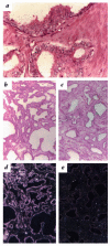
Downregulation of prostatic VEGF mRNA expression by androgen-ablation therapy. (a) In situ hybridization of a neoplastic prostate specimen with a VEGF-specific probe shown at high magnification. Note abundant expression of VEGF in the abnormal glandular epithelium. (be) Low-power magnifications to show global changes in VEGF expression via in situ hybridization of grade-matched specimens either untreated (b, bright-field; d, dark-field) or subjected to androgen-ablation treatment before prostatectomy (c, bright-field; e, dark-field). Sections were cohybridized on the same slide. Note a marked reduction in VEGF expression as a result of androgen-ablation therapy.
Figure 4
Figure 4
Androgen-ablation therapy in prostate cancer leads to selective obliteration of immature vessels. Adjacent sections of surgically removed prostate tissues were immunostained for vWF (a and c) or for α-SMA (b and d) to examine individual vessels for coverage with periendothelial cells. One example is shown for an untreated tumor (a and b) (black arrows pointing at uncovered vessels and a blue arrow pointing at a covered vessel) and one for a tumor subjected to androgen-ablation therapy (c and d) (black arrows pointing at a covered vessel). Data from 10 different patients are shown in the histogram (e): five from control untreated tumors (hatched bars) and five from treated tumors (solid bars). To randomize for experimental variability during processing and immunohistochemical detection, pairs of tumors, each containing one control tumor and an androgen-ablated tumor of the same Gleason grade and a matching patient age, were embedded in a single block and coanalyzed on the same slide. The total number of lumenized vessels scored depended on the amount of tumor represented in the section and was as follows: tumor 1 (140), 2 (63), 3 (25), 4 (19), 5 (98), 6 (122), 7 (158), 8 (143), 9 (170), 10 (397). Mouse prostate was used as a control for vascular maturation in normal prostate. On average, untreated tumors contained 38% α-SMA–positive vessels (SEM = 3.5%); androgen-ablated tumors had 79% α-SMA–positive vessels (SEM = 3.3%).
Figure 5
Figure 5
Endothelial cell apoptosis after androgen ablation. TUNEL analysis was used to detect apoptotic cells in the untreated prostate and 4 weeks after hormone ablation. (a) An untreated prostate showing apoptotic nuclei (red) in glands but not in blood vessels (arrows). (b) An androgen-ablated specimen highlighting two blood vessels with several TUNEL-positive endothelial cells. Note the presence of TUNEL-positive (black arrow) and TUNEL negative (red arrow) in the same blood vessel. a and b were processed together on the same microscope slide to control for histochemical variability. (c) TUNEL (green fluorescence) and α-SMA staining (red fluorescence) showing that an uncovered blood vessel (arrowhead) contains many apoptotic endothelial cells, whereas an adjacent covered blood vessel (arrow) does not. Red autofluorescence of erythrocytes aids in identifying the lumen of these vessels. Eighty-five percent of vessels in which one or more TUNEL-positive endothelial cells were detected were α-SMA–negative. The mean number of TUNEL-positive endothelial cells per vessel was 3.1-fold greater in androgen-ablated tumors. TUNEL, terminal deoxynucleotide transferase–mediated dUTP nick end-labeling.

Comment in

References

    1. Folkman, J. 1997. Cancer: principles and practice of oncology. Lippincott-Raven Publishers. Philadelphia, PA. 3075–3085.
    1. Holmgren L, O’Reilly MS, Folkman J. Dormancy of micrometastases: balanced proliferation and apoptosis in the presence of angiogenesis suppression. Nat Med. 1995;1:149–153. - PubMed
    1. Millauer B, et al. Dominant-negative inhibition of Flk-1 suppresses the growth of many tumor types in vivo. Cancer Res. 1996;56:1615–1620. - PubMed
    1. Warren RS, Yuan H, Matli MR, Gillett NA, Ferrara N. Regulation by vascular endothelial growth factor of human colon cancer tumorigenesis in a mouse model of experimental liver metastasis. J Clin Invest. 1995;95:1789–1797. - PMC - PubMed
    1. Borgstrom P, Hillan KJ, Sriramarao P, Ferrara N. Complete inhibition of angiogenesis and growth of microtumors by anti-vascular endothelial growth factor neutralizing antibody: novel concepts of angiostatic therapy from intravital videomicroscopy. Cancer Res. 1996;56:4032–4039. - PubMed

Publication types

MeSH terms