. 1999 Jan;103(2):185-95.
doi: 10.1172/JCI3326.

Glucose or diabetes activates p38 mitogen-activated protein kinase via different pathways

Affiliations

Glucose or diabetes activates p38 mitogen-activated protein kinase via different pathways

M Igarashi et al. J Clin Invest. 1999 Jan.

Abstract

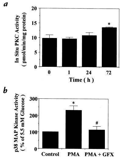
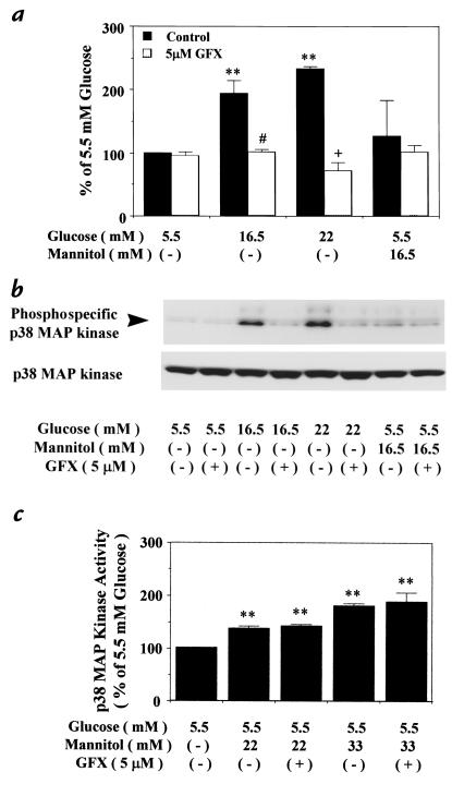
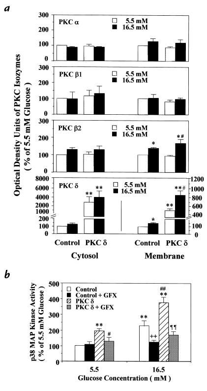
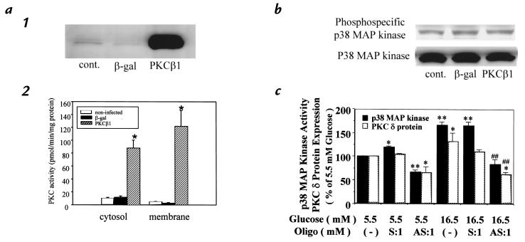
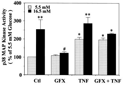
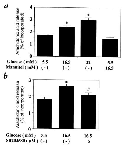
Hyperglycemia can cause vascular dysfunctions by multiple factors including hyperosmolarity, oxidant formation, and protein kinase C (PKC) activation. We have characterized the effect of hyperglycemia on p38 mitogen-activated protein (p38) kinase activation, which can be induced by oxidants, hyperosmolarity, and proinflammatory cytokines, leading to apoptosis, cell growth, and gene regulation. Glucose at 16.5 mM increased p38 kinase activity in a time-dependent manner compared with 5.5 mM in rat aortic smooth muscle cells (SMC). Mannitol activated p38 kinase only at or greater than 22 mM. High glucose levels and a PKC agonist activated p38 kinase, and a PKC inhibitor, GF109203X, prevented its activation. However, p38 kinase activation by mannitol or tumor necrosis factor-alpha was not inhibited by GF109203X. Changes in PKC isoform distribution after exposure to 16.5 mM glucose in SMC suggested that both PKC-beta2 and PKC-delta isoforms were increased. Activities of p38 kinase in PKC-delta- but not PKC-beta1-overexpressed SMC were increased compared with control cells. Activation of p38 kinase was also observed and characterized in various vascular cells in culture and aorta from diabetic rats. Thus, moderate hyperglycemia can activate p38 kinase by a PKC-delta isoform-dependent pathway, but glucose at extremely elevated levels can also activate p38 kinase by hyperosmolarity via a PKC-independent pathway.


Figures

Figure 1
Figure 1
(a) Effect of glucose on ERK-1, ERK-2, and p38 MAP kinase activations in rat aortic SMC. After 72 h of exposure to 5.5, 16.5, or 22 mM glucose, the cells were lysed, and MAP kinase activities were quantitated by in vitro phosphorylation of MBP using [γ-32P]ATP as described in Methods. The results were derived from three separate experiments, with duplicates performed in each experiment. Each bar represents the mean ± SEM in a, b, and c. *P < 0.05, **P < 0.01 vs. 5.5 mM glucose. (b) Time course of 16.5 mM glucose on p38 MAP kinase activity. After the indicated time of incubation with 16.5 mM glucose, the cells were lysed and p38 MAP kinase activity was quantitated as already described. The results were derived from four separate experiments, with duplicates performed in each experiment. *P < 0.05, **P < 0.01 vs. 0 h. (c) Effect of elevated glucose and mannitol levels on p38 MAP kinase activation. After 72 h of exposure to several concentrations of glucose or mannitol, the cells were lysed and p38 MAP kinase activity was quantitated as already described. The results were derived from three separate experiments, with duplicates performed in each experiment. *P < 0.05, **P < 0.01 vs. 5.5 mM glucose; #P < 0.05 vs. 11 mM glucose; ++P < 0.01 vs. 16.5 mM glucose; P < 0.05 vs. 22 mM glucose. ERK, extracellular signal-regulated protein kinase; MAP, mitogen-activated protein; MBP, myelin basic protein; SMC, smooth muscle cells.
Figure 2
Figure 2
(a) Time course of glucose's 16.5 mM effect on in situ PKC activity in rat aortic SMC. After indicated time of incubation with 16.5 mM glucose, PKC activities were measured by in situ PKC assay using a PKC-specific peptide substrate, RKRTLRRL, in digitonin-permeabilized cells as described in Methods. *P < 0.05 vs. 0 h. (b) Effect of regulators of PKC on p38 MAP kinase activation. After subculturing with 5.5 mM glucose, the cells were treated or not treated with PMA (100 nM) for 15 min or DMSO, treated with a PKC-specific inhibitor, GF109203X (GFX, 5 μM) pr ethanol, for 30 min, and then lysed. p38 MAP kinase activity was quantitated by in vitro phosphorylation of MBP using [γ-32P]ATP as described in Methods. GF109203X was solubilized with DMSO and PMA with ethanol, respectively, with final concentrations of DMSO and ethanol of 0.1%. The results were derived from three separate experiments. *P < 0.05 vs. 5.5 mM glucose (–), #P < 0.05 vs. 5.5 mM glucose treated with PMA. Each bar represents the mean ± SEM. PKC, protein kinase C.
Figure 3
Figure 3
Effect of GF109203X on p38 MAP kinase activation in rat aortic SMC. After 72 h of exposure to several concentrations of glucose or mannitol, the cells were untreated, or treated with a PKC-specific inhibitor, GF109203X (GFX, 5 μM), for 30 min; then, the cells were lysed as described in Methods. (a) p38 MAP kinase activity measured by the phosphorylation of MBP using [γ-32P]ATP. The results were derived from four separate experiments. **P < 0.01 vs. 5.5 mM glucose, #P < 0.05 vs. 16.5 mM glucose, +P < 0.05 vs. 22 mM glucose. (b) Expression of p38 MAP kinase by immunoblot analysis. The samples were separated by 10% SDS-PAGE, transferred to PVDF membranes, and blocked overnight. The membranes were incubated with antiphosphospecific p38 MAP kinase and anti-p38 antibody. The arrowhead shows the phosphorylation of p38 MAP kinase. The results were derived from three separate experiments. (c) p38 MAP kinase activity by the phosphorylation of MBP using [γ-32P]ATP. The results were derived from three separate experiments, with each experiment performed in duplicate. **P < 0.01 vs. 5.5 mM glucose. Each bar represents the mean ± SEM. PVDF, polyvinyldene difluoride.
Figure 4
Figure 4
Effect of a PKC-β–specific inhibitor, LY333351, on p38 MAP kinase activity in rat aortic SMC. After 3 days of exposure to several concentrations of glucose or mannitol, the cells were untreated or treated with a PKC-specific inhibitor, LY333351 [(a) 20 nM and (b) 500 nM], for 30 min, and then lysed. The p38 MAP kinase activity was quantitated by the phosphorylation of MBP using [γ-32P]ATP as described in Methods. LY333351 was solubilized with DMSO. The results were derived from four separate experiments, with each experiment performed in duplicate. Each bar represents the mean ± SEM. *P < 0.05 vs. 5.5 mM glucose.
Figure 5
Figure 5
(a) Characterization of PKC isoform and activities in PKC-δ isoform–overexpressed cells. (b) Immunoblot analysis of PKC isoforms in PKC-δ−overexpressed and the control cells containing the retroviral vector without PKC-δ cDNA derived from rat aortic SMC. The infection was performed using the retroviral vector pBabe-Puro, as described in Methods. After the vector with or without PKC-δ, cDNA was transfected into BOSC23 cells via Lipofectamine, and the supernatant was filtered and added to primary cultured rat SMC for infection. Positive transfectants were selected using puromysin. PKC proteins in both membranous and cytosolic fractions were partially purified. The proteins were separated by 8% SDS-PAGE, and immunoblot analysis was performed using various types of antibodies on PKC isoforms as described in Methods. The results are derived from four separate experiments. Each bar represents the mean ± SEM. *P < 0.05, **P < 0.01 vs. control cells at 5.5 mM glucose; #P < 0.05 vs. PKC-δ−overexpressed cells at 5.5 mM glucose. (b) Effect of PKC-δ overexpression on p38 MAP kinase activity in rat aortic SMC. After 72 h of exposure to 5.5 mM or 16.5 mM glucose, the cells were untreated or treated with a PKC-specific inhibitor, GF109203X (GFX, 5 μM), for 30 min, and then lysed. The p38 MAP kinase activity was quantitated by the phosphorylation of MBP using [γ-32P]ATP as described in Methods. The results were derived from four separate experiments, with each experiment performed in duplicate. Each bar represents the mean ± SEM. **P < 0.01 vs. control at 5.5 mM glucose; #P < 0.05, ##P < 0.01 vs. PKC-δ–overexpressed cells at 5.5 mM glucose; ++P < 0.01 vs. control at 16.5 mM glucose; ¶¶P < 0.01 vs. PKC-δ–overexpressed cells at 16.5 mM glucose. These results were derived from three different cloned populations of SMC overexpressing PKC-δ.
Figure 6
Figure 6
Effect of PKC-β1 overexpression on p38 MAP kinase in rat aortic SMC. (a) Characterization of PKC-β1 overexpression in rat aortic SMC after adenovirus infection. (1) Total cell lysate was isolated from cells that were not infected (cont.), or infected with Adv-CMV-βGal (β-gal) or Adv-CMV-PKC-β1 (PKC-β1). PKC-β1 protein was detected by immunoblot analysis. (2) Four days after infection with adenovirus, cells were lysed and the cytosol and membrane proteins were fractionated by ultracentrifugation. The PKC activity was measured as described in Methods. Each bar represents the mean + SD (n = 5). *P < 0.001 vs. noninfected and β-gal. (b) Activation of p38 MAP kinase in PKC-β1–overexpressed SMC. Four days after infection with adenovirus, total cell lysate was isolated. The samples were separated by 10% SDS-gel, and the phosphorylation of p38 MAP kinase was detected by immunoblot analysis using phosphospecific p38 MAP kinase as described in Methods. The results were derived from three separate experiments.
Figure 7
Figure 7
Effect of TNF-α and GF109203X on p38 MAP kinase activity in rat aortic SMC. After subculturing with 5.5 mM or 16.5 mM glucose, the cells were not treated or treated with TNF-α (100 nM) for 5 min after pretreatment with a PKC-specific inhibitor, GF109203X (GFX, 5 μM), for 30 min, and then lysed. The p38 MAP kinase activity was quantitated by the phosphorylation of MBP using [γ-32P]ATP as described in Methods. GF109203X was solubilized with DMSO and TNF-α with serum-free DMEM, respectively. The results were derived from three separate experiments, with each experiment performed in duplicate. Each bar represents the mean ± SEM. *P < 0.05, **P < 0.01 vs. 5.5 mM glucose (–); #P < 0.05 vs. 16.5 mM glucose (–). TNF, tumor necrosis factor.
Figure 8
Figure 8
Effect of elevated glucose levels and a p38 MAP kinase–specific inhibitor, SB-203580, on arachidonic acid release in rat aortic SMC. (a) After 48 h of exposure to various conditions of glucose and mannitol, the cells were labeled with [3H]arachidonic acid and further subcultured with the same conditions. The media were collected, and the amount of [3H]arachidonic acid released into the media was measured as described in Methods. Each bar represents the mean ± SEM. *P < 0.05 vs. 5.5 mM glucose. (b) SB-203580 was added to 0.5 ml of the media and incubated for 30 min before the labeled media were collected for measurement of arachidonic acid release. Each bar represents the mean ± SEM. *P < 0.01 vs. 5.5 mM glucose (–), #P < 0.05 vs. 16.5 mM glucose.
Figure 9
Figure 9
Effect of hyperglycemia on p38 MAP kinase activation in the aorta derived from control and diabetic rats. Rats were treated as described in Methods, and expressions of p38 MAP kinase and phosphospecific p38 MAP kinase were performed by immunoblot analysis. The optical density units by immunoblot analysis were measured with an image densitometer. Each bar represents the mean ± SEM (n = 8). **P < 0.01 vs. the control.

References

    1. The Diabetes ControlComplications Trial Research Group. The effect of intensive treatment of diabetes on the development and progression of long-term complications in insulin dependent diabetes mellitus. N Engl J Med. 1993;329:977–986. - PubMed
    1. Greene D, Lattimer SA, Sima AAF. Sorbitol, phosphoinositides and sodium-potassium-ATPase in the pathogenesis of diabetic complications. N Engl J Med. 1987;316:599–606. - PubMed
    1. Brownlee M, Cerami A, Vlassara H. Advanced glycosylation end products in tissue and biochemical basis of diabetic complications. N Engl J Med. 1988;318:1315–1321. - PubMed
    1. Williamson JR, et al. Hyperglycemic pseudhypoxia and diabetic complications. Diabetes. 1993;42:801–813. - PubMed
    1. Koya D, King GL. Protein kinase C activation and the development of diabetic complication. Diabetes. 1998;47:859–866. - PubMed

Publication types

MeSH terms