. 1999 Jan;103(2):197-206.
doi: 10.1172/JCI3523.

TGF-beta signaling blockade inhibits PTHrP secretion by breast cancer cells and bone metastases development

Affiliations

TGF-beta signaling blockade inhibits PTHrP secretion by breast cancer cells and bone metastases development

J J Yin et al. J Clin Invest. 1999 Jan.

Abstract

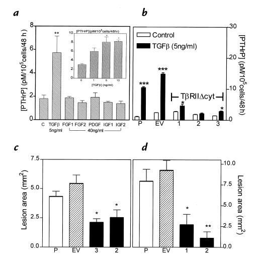
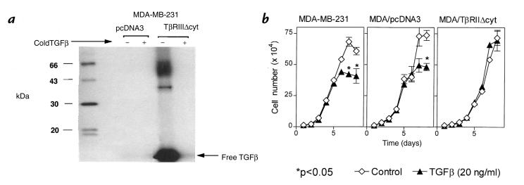
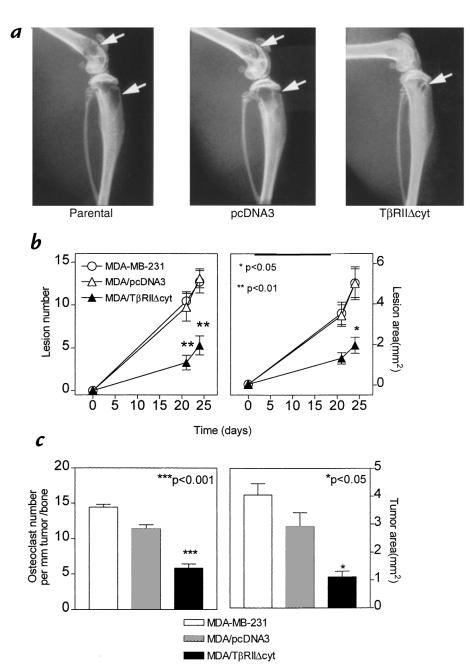
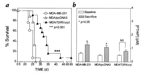
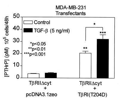
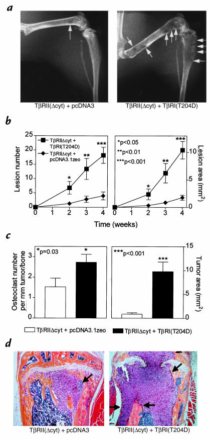
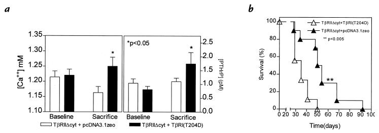
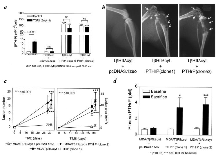
Breast cancer frequently metastasizes to the skeleton, and the associated bone destruction is mediated by the osteoclast. Growth factors, including transforming growth factor-beta (TGF-beta), released from bone matrix by the action of osteoclasts, may foster metastatic growth. Because TGF-beta inhibits growth of epithelial cells, and carcinoma cells are often defective in TGF-beta responses, any role of TGF-beta in metastasis is likely to be mediated by effects on the surrounding normal tissue. However, we present evidence that TGF-beta promotes breast cancer metastasis by acting directly on the tumor cells. Expression of a dominant-negative mutant (TbetaRIIDeltacyt) of the TGF-beta type II receptor rendered the human breast cancer cell line MDA-MB-231 unresponsive to TGF-beta. In a murine model of bone metastases, expression of TbetaRIIDeltacyt by MDA-MB-231 resulted in less bone destruction, less tumor with fewer associated osteoclasts, and prolonged survival compared with controls. Reversal of the dominant-negative signaling blockade by expression of a constitutively active TGF-beta type I receptor in the breast cancer cells increased tumor production of parathyroid hormone-related protein (PTHrP), enhanced osteolytic bone metastasis, and decreased survival. Transfection of MDA-MB-231 cells that expressed the dominant-negative TbetaRIIDeltacyt with the cDNA for PTHrP resulted in constitutive tumor PTHrP production and accelerated bone metastases. These data demonstrate an important role for TGF-beta in the development of breast cancer metastasis to bone, via the TGF-beta receptor-mediated signaling pathway in tumor cells, and suggest that the bone destruction is mediated by PTHrP.


Figures

Figure 1
Figure 1
(a) Effects of bone growth factors on PTHrP secretion from MDA-MB-231 cells in vitro. MDA-MB-231 cells were plated onto 48-well plates and grown to near confluence. Cells were washed and treated with serum-free media containing the respective growth factors for 48 h. PTHrP concentrations in conditioned media were corrected for cell number. Only the results for the highest concentration of each growth factor are shown. Inset: Dose response for PTHrP secretion by MDA-MB-231 cells treated with TGF-β. Values represent the mean ± SEM (n = 3 per group).(b) Effect of TGF-β on PTHrP secretion by MDA-MB-231, MDA/pcDNA3, and MDA/TβRIIΔcyt clones. Respective cells were plated onto 48-well plates and treated as described in a. Values represent the mean ± SEM (n = 3 per group). P = parental MDA-MB-231; EV = empty vector pcDNA3 clone; 1, 2, and 3 are respective MDA/TβRIIΔcyt clones. (c and d) Osteolytic lesion area from radiographs of two separate experiments comparing clones 3 and 2 (c) or clones 1 and 2 (d) with controls of MDA-MB-231 (P) or pcDNA3 vector (EV). Values represent mean ± SEM (n = 4 per group). *P < 0.05, **P < 0.01, ***P < 0.001 vs. controls. PTHrP, parathyroid hormone–related protein; TGF-β, transforming growth factor-β.
Figure 2
Figure 2
(a) 125I-labeled TGF-β1 cross-linking followed by immunoprecipitation with anti-HA antibody in clonal MDA-MB-231 cells expressing the TβRIIΔcyt (MDA/TβRIIΔcyt) or the empty vector (MDA/pcDNA3). Clonal lines were cross-linked with 160 pM 125I-labeled TGF-β1 alone (–) or in the presence of excess unlabeled TGF-β1 (5 nM) (+). Cell extracts were subjected to SDS-PAGE and autoradiography to visualize the truncated receptor. (b) Effect of TGF-β on growth rate of MDA-MB-231, MDA/pcDNA3, and MDA/TβRIIΔcyt cells. Respective cells were plated at a density of 104 cells per well with or without TGF-β (20 ng/ml) in 10% FCS and counted daily. Values represent the mean ± SEM (n = 3 per group). In the absence of TGF-β, growth rates of each cell line did not significantly differ.
Figure 3
Figure 3
(a) Representative radiographs of hindlimbs from mice bearing MDA-MB-231, MDA/pcDNA3, or MDA/TβRIIΔcyt tumors 24 days after tumor inoculation. Osteolytic lesions are indicated by the arrows. (b) Osteolytic lesion number and area on radiographs as measured by computerized image analysis of forelimbs and hindlimbs. Respective tumor cells were inoculated on day 0. Values represent the mean ± SEM (n = 13 per group). (c) Histomorphometric analysis of forelimbs and hindlimbs from mice with osteolytic lesions. Data represent measurements from midsections of tibiae, femora, and humeri of mice from b, inoculated with either MDA-MB-231, MDA/pcDNA3, or MDA/TβRIIΔcyt clone 2. Tumor area (mm2) from metastatic bone lesions is illustrated on the right and osteoclast number per millimeter of tumor adjacent to bone (tumor/bone interface) on the left. Values represent the mean ± SEM.
Figure 4
Figure 4
Survival (a) and plasma PTHrP concentrations (b) in mice bearing MDA-MB-231, MDA/pcDNA3, or MDA/TβRIIΔcyt tumors. (a) Survival of mice bearing MDA/TΔRIIΔcyt tumors was significantly longer than that of the controls. (b) Plasma PTHrP concentrations at sacrifice were significantly higher than respective concentrations before tumor inoculation (baseline) in mice bearing control tumors of MDA-MB-231 or MDA/pcDNA3. There was no significant difference between baseline and sacrifice values in mice bearing the MDA/TβRIIΔcyt tumors.
Figure 5
Figure 5
Effect of TGF-β on PTHrP secretion by MDA-MB-231 clonal lines, TβRIIΔcyt + TβRI(T204D), and TβRIIΔcyt + pcDNA3.1zeo. Respective cells were plated onto 48-well plates and treated as in Fig. 1a. Values represent the mean ± SEM (n = 3 per group). **P < 0.01 and ***P < 0.001 compared with TβRIIΔcyt + pcDNA3.1zeo (control or TGF-β–stimulated).
Figure 6
Figure 6
(a) Representative radiographs of hindlimbs from mice bearing TβRIIΔcyt + TβRI(T204D) or TβRIIΔcyt + pcDNA3.1zeo 28 days after tumor inoculation. Osteolytic lesions are indicated by the arrows. (b) Osteolytic lesion number and area on radiographs as measured by computerized image analysis of forelimbs and hindlimbs. Respective tumor cells were inoculated on day 0. Values represent the mean ± SEM (n = 5 per group). (c) Histomorphometric analysis of hindlimbs from mice with osteolytic lesions. Data represent measurements from midsections of tibiae and femora of mice (from b) inoculated with either TβRIIΔcyt + TβRI(T204D) or TβRIIΔcyt + pcDNA3.1zeo tumors. Tumor area (mm2) from metastatic bone lesions is illustrated on the right and osteoclast number per millimeter of tumor adjacent to bone (tumor/bone interface) on the left. Values represent the mean ± SEM. (d) Bone histology from the midtibial metaphysis of representative mice bearing either TβRIIΔcyt + TβRI(T204D) or TβRIIΔcyt + pcDNA3.1zeo tumors. Tumor (arrows) filled the marrow cavity and replaced normal cellular elements in mice bearing TβRIIΔcyt + TβRI(T204D) tumors (right). There was significant loss of both cortical and trabecular bone in this group, and tumor has eroded through the growth plate. In contrast, sections from mice bearing control TβRIIΔcyt + pcDNA3.1zeo tumors (left) had small foci of tumor in the marrow cavity (arrows) with little bone destruction, as evidenced by intact trabecular and cortical bone.
Figure 7
Figure 7
(a) Plasma PTHrP concentrations (right) and whole blood ionized calcium concentrations (left) at sacrifice were significantly higher than respective concentrations before tumor inoculation (baseline) in mice bearing tumors TβRIIΔcyt + TβRI(T204D). There was no significant difference between baseline and sacrifice values in mice bearing the control TβRIIΔcyt + pcDNA3.1zeo tumors. (b) Survival of mice bearing TβRIIΔcyt + TβRI(T204D) was significantly shorter than that of the controls, TβRIIΔcyt + pcDNA3.1zeo (n = 10 per group).
Figure 8
Figure 8
(a) Effect of TGF-β on PTHrP secretion by MDA-MB-231 and MDA/TβRIIΔcyt cell clones that overexpress PTHrP (TβRIIΔcyt + PTHrP; two clones) or the empty vector (TβRIIΔcyt + pcDNA3.1zeo). Respective cells were plated onto 48-well plates and treated as in Fig. 1a. Values represent the mean ± SEM (n = 3 per group). (b) Representative radiographs of hindlimbs from mice bearing two different TβRIIΔcyt + PTHrP clones or TβRIIΔcyt + pcDNA3.1zeo control 31 days after tumor inoculation. Osteolytic lesions are indicated by the arrows. (c) Osteolytic lesion number and area on radiographs as measured by computerized image analysis of forelimbs and hindlimbs. Respective tumor cells were inoculated on day 0. Values represent the mean ± SEM (n = 5) per group. (d) Plasma PTHrP concentrations at sacrifice were significantly higher than respective concentrations prior to tumor inoculation (baseline) in mice bearing either TβRIIΔcyt + PTHrP tumors. There was no significant difference between baseline and sacrifice values in mice bearing the control TβRIIΔcyt + pcDNA3.1zeo tumors.

References

    1. Paget S. The distribution of secondary growths in cancer of the breast. Lancet. 1889;1:571–572. - PubMed
    1. Coleman RE, Rubens RD. The clinical course of bone metastases from breast cancer. Br J Cancer. 1987;55:61–66. - PMC - PubMed
    1. Hauschka PV, Mavrakos AE, Iafrati MD, Doleman SE, Klagsbrun M. Growth factors in bone matrix. J Biol Chem. 1986;261:12665–12674. - PubMed
    1. Pfeilschifter J, Mundy GR. Modulation of transforming growth factor β activity in bone cultures by osteotrophic hormones. Proc Natl Acad Sci USA. 1987;84:2024–2028. - PMC - PubMed
    1. Boyde A, Maconnachie E, Reid SA, Delling G, Mundy GR. Scanning electron microscopy in bone pathology: review of methods. Potential and applications. Scanning Electron Microsc. 1986;4:1537–1554. - PubMed

Publication types

MeSH terms

Substances