. 1999 Jan;154(1):61-6.
doi: 10.1016/S0002-9440(10)65251-0.

Immuno-LCM: laser capture microdissection of immunostained frozen sections for mRNA analysis

Affiliations

Immuno-LCM: laser capture microdissection of immunostained frozen sections for mRNA analysis

F Fend et al. Am J Pathol. 1999 Jan.

Abstract

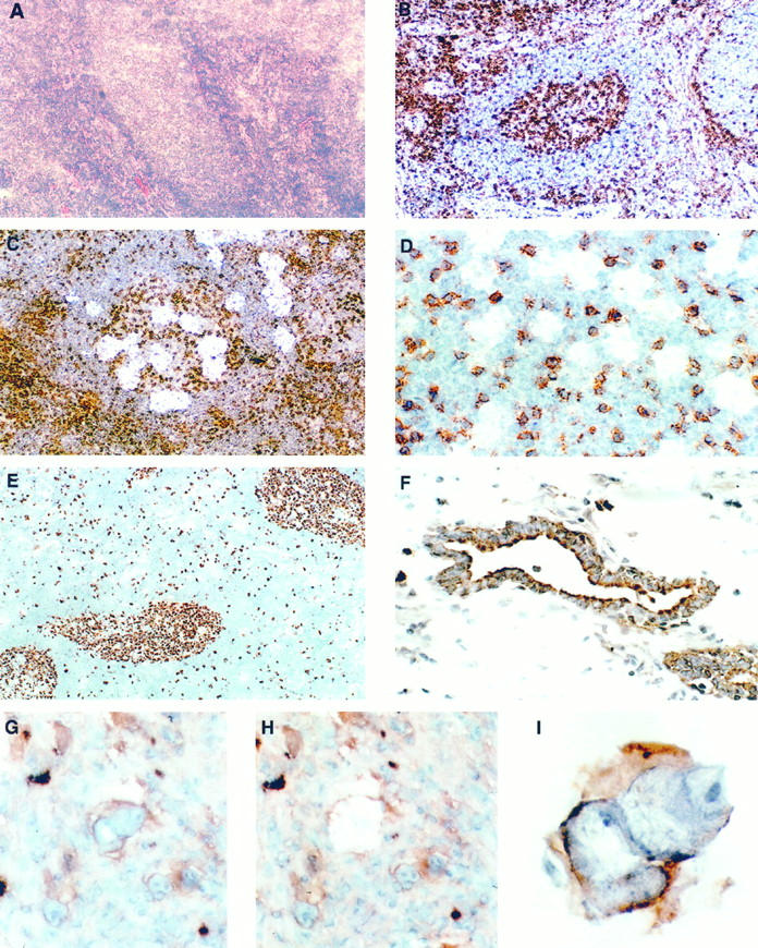
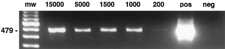
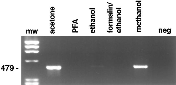
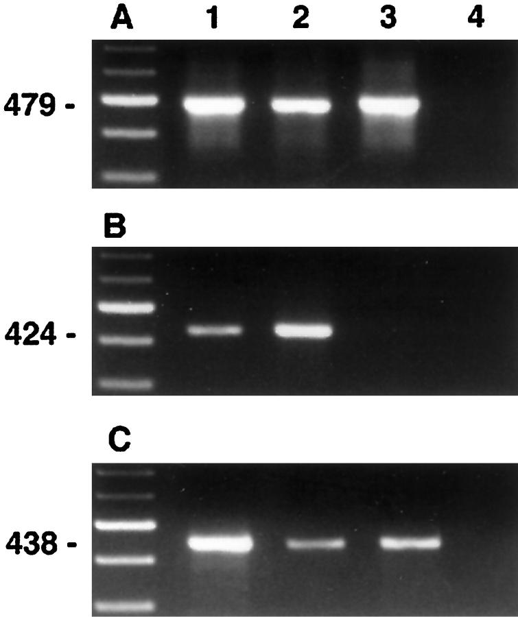
Microdissection of routinely stained or unstained frozen sections has been used successfully to obtain purified cell populations for the analysis of cell-specific gene expression patterns in primary tissues with a complex mixture of cell types. However, the precision and usefulness of microdissection is frequently limited by the difficulty to identify different cell types and structures by morphology alone. We therefore developed a rapid immunostaining procedure for frozen sections followed by laser capture microdissection (LCM) and RNA extraction, which allows targeted mRNA analysis of immunophenotypically defined cell populations. After fixation, frozen sections are immunostained under RNAse-free conditions using a rapid three-step streptavidin-biotin technique, dehydrated and immediately subjected to LCM. RNA is extracted from captured tissue, DNAse I treated, and reverse transcribed. Acetone-, methanol-, or ethanol/acetone-fixed sections give excellent immunostaining after 12 to 25 minutes total processing time. Specificity, precision, and speed of microdissection is markedly increased due to improved identification of desired (or undesired) cell types. The mRNA recovered from immunostained tissue is of high quality. Single-step PCR is able to amplify fragments of more than 600 bp from both housekeeping genes such as beta-actin as well as cell-specific messages such as CD4 or CD19, using cDNA derived from less than 500 immunostained, microdissected cells. Immuno-LCM allows specific mRNA analysis of cell populations isolated according to their immunophenotype or expression of function-related antigens and significantly expands our ability to investigate gene expression in heterogeneous tissues.


Figures

Figure 1.
Figure 1.
Frozen sections of a reactive lymph node with a central follicle stained with H&E (A) and immunostained for CD45RO (B to D). The identification of the follicle is difficult in the routinely stained section, whereas immunostaining highlights T cells within the germinal center and in the perifollicular area and makes architectural features more evident. Magnification, ×100. Microdissection from germinal centers with spot sizes of 50 to 60 μm (C) and 15 to 20 μm (D) in diameter as seen during LCM, without coverslip. The smaller spot size allows avoidance of interspersed T cells. Magnification, ×100 and ×400. E: Lymph node stained for the proliferation-associated antigen Ki67, using MIB1 antibody (dilution, 1:10) in conjunction with the Quickstain kit. Magnification, ×100. F: Prostate immunostained for cytokeratin. Stromal tissue has been removed by LCM, leaving behind the positively stained glands. Magnification, ×400. G to I: Microdissection of a single Reed-Sternberg cell stained for CD30 before (G) and after (H) removal and the isolated cell adherent to the membrane (I). Magnification, ×400 and ×1000.
Figure 2.
Figure 2.
RT-PCR of a 479-bp β-actin fragment from microdissected tissue fragments obtained from an immunostained lymph node after acetone fixation. The estimated total number of dissected cells is indicated above the lane. Approximately 1/20 of the total cDNA volume obtained was used per reaction. Forty cycles of amplification were used.
Figure 3.
Figure 3.
RT-PCR amplification of a 479-bp β-actin fragment from immunostained frozen sections of a reactive lymph node showing the influence of different fixatives on RNA recovery. Lane 1, molecular weight marker; lane 2, acetone; lane 3, 4% paraformaldehyde; lane 4, 70% ethanol; lane 5, formalin/ethanol; lane 6, methanol; lane 7, water control. Thirty-five cycles of amplification were used.
Figure 4.
Figure 4.
RT-PCR amplification of β-actin (A), CD19 (B), and CD4 (C) from a reactive lymph node immunostained for CD45RO. Microdissectates were obtained randomly (lane 1), from B cell areas (lane 2), and T cell areas (lane 3). Lane 4, negative control. The weak CD4 signal in lane 2 probably results from contaminating T cells or CD4-expressing macrophages not identified on the CD45RO stain.

References

    1. Whetsell L, Maw G, Nadon N, Ringer PD, Schaefer FV: Polymerase chain reaction microanalysis of tumors from stained histological slides. Oncogene 1992, 7:2355-2361 - PubMed
    1. Going JJ, Lamb RF: Practical histological microdissection for PCR analysis. J Pathol 1996, 179:121-124 - PubMed
    1. Emmert-Buck MR, Bonner RF, Smith PD, Chuaqui R, Zhuang Z, Goldstein SR, Weiss RA, Liotta LA: Laser capture microdissection. Science 1996, 274:998-1001 - PubMed
    1. Böhm M, Wieland I, Schütze K, Rübben H: Microbeam MOMenT: non-contact laser microdissection of membrane-mounted native tissue. Am J Pathol 1997, 151:63-67 - PMC - PubMed
    1. Shibata D, Hawes D, Li Z-H, Hernandez AM, Spruck CH, Nichols PW: Specific genetic analysis of microscopic tissue after selective ultraviolet radiation fractionation and the polymerase chain reaction. Am J Pathol 1992, 141:539-543 - PMC - PubMed

Publication types