. 1999 Feb 1;19(3):964-73.
doi: 10.1523/JNEUROSCI.19-03-00964.1999.

Mutant huntingtin expression in clonal striatal cells: dissociation of inclusion formation and neuronal survival by caspase inhibition

Affiliations

Mutant huntingtin expression in clonal striatal cells: dissociation of inclusion formation and neuronal survival by caspase inhibition

M Kim et al. J Neurosci. .

Abstract

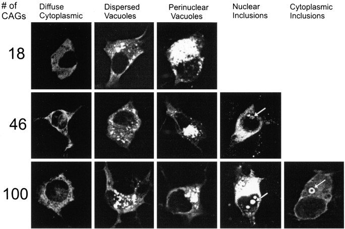
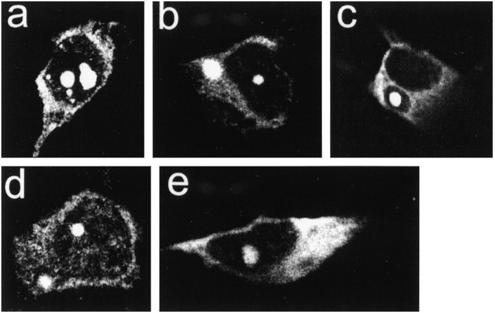
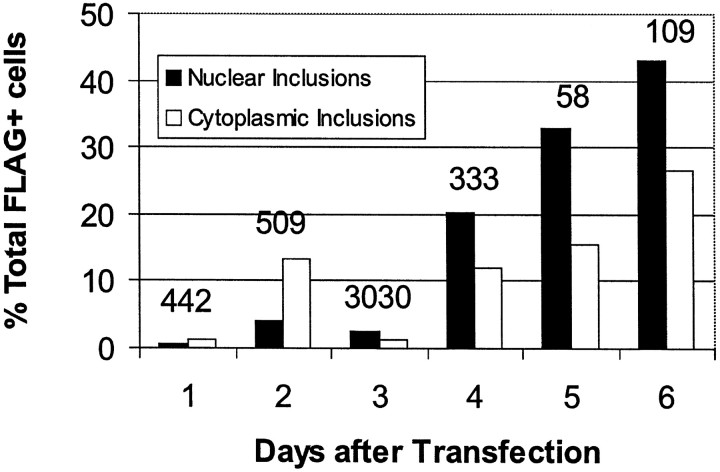
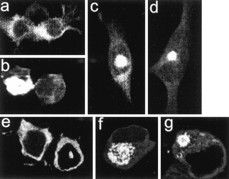
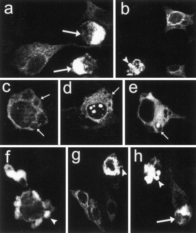
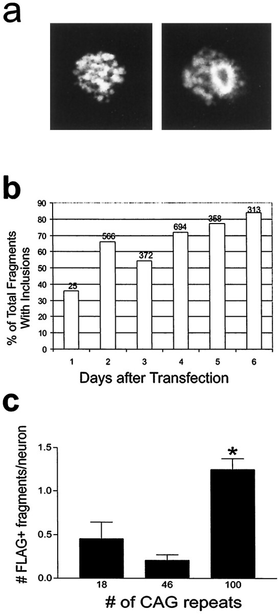
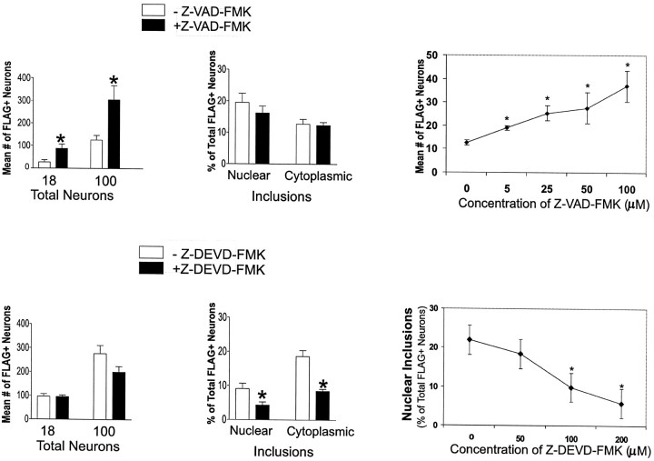
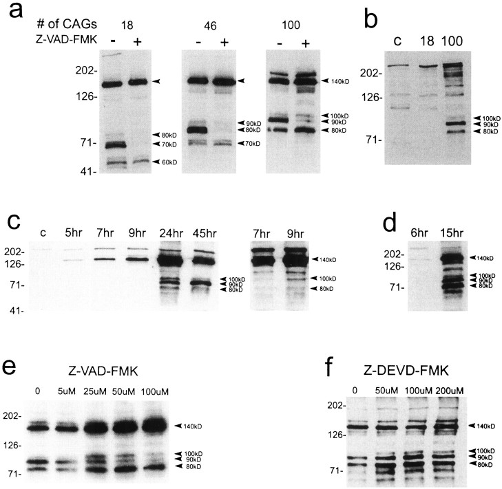
Neuronal intranuclear inclusions are found in the brains of patients with Huntington's disease and form from the polyglutamine-expanded N-terminal region of mutant huntingtin. To explore the properties of inclusions and their involvement in cell death, mouse clonal striatal cells were transiently transfected with truncated and full-length human wild-type and mutant huntingtin cDNAs. Both normal and mutant proteins localized in the cytoplasm, and infrequently, in dispersed and perinuclear vacuoles. Only mutant huntingtin formed nuclear and cytoplasmic inclusions, which increased with polyglutamine expansion and with time after transfection. Nuclear inclusions contained primarily cleaved N-terminal products, whereas cytoplasmic inclusions contained cleaved and larger intact proteins. Cells with wild-type or mutant protein had distinct apoptotic features (membrane blebbing, shrinkage, cellular fragmentation), but those with mutant huntingtin generated the most cell fragments (apoptotic bodies). The caspase inhibitor Z-VAD-FMK significantly increased cell survival but did not diminish nuclear and cytoplasmic inclusions. In contrast, Z-DEVD-FMK significantly reduced nuclear and cytoplasmic inclusions but did not increase survival. A series of N-terminal products was formed from truncated normal and mutant proteins and from full-length mutant huntingtin but not from full-length wild-type huntingtin. One prominent N-terminal product was blocked by Z-VAD-FMK. In summary, the formation of inclusions in clonal striatal cells corresponds to that seen in the HD brain and is separable from events that regulate cell death. N-terminal cleavage may be linked to mutant huntingtin's role in cell death.


Figures

Fig. 1.
Fig. 1.
Patterns of localization of FLAG–huntingtin fusion proteins in clonal striatal neurons 1–3 d after transfection of FH3221 with 18, 46, or 100 glutamines. Diffuse cytoplasmic labeling, dispersed vacuoles, and condensed perinuclear vacuoles occur regardless of polyglutamine length. Neurons with mutant huntingtin show some diffuse nuclear labeling and nuclear and cytoplasmic (“ring-like”) inclusions (arrows).
Fig. 2.
Fig. 2.
Features of NI and CI in clonal striatal and midbrain neurons. The inclusions were formed by FH3221-100 (a–c, e) or FH9774-100 (d). Inclusions in striatal cells appear more consolidated and intensely FLAG-positive (a–d) than in the midbrain neuron (e). Striatal cell ina has three small and two large NI. Cells inb and d have both a nuclear and a cytoplasmic inclusion. Cell in c shows retraction of cytoplasm away from the cytoplasmic inclusion. All cells have diffuse cytoplasmic labeling, and cell in d has diffuse nuclear labeling.
Fig. 3.
Fig. 3.
Effects of the number of days after transfection on the formation of NI and CI. Cells were transfected with FH3221-100. Percent of total FLAG+ neurons with inclusions progressively increases between 4 and 6 d. Note that the total number of FLAG+ neurons (indicated over each set of bars) decreases markedly by 5–6 d after transfection. Results are shown for one transfection experiment. Results were similar in two other experiments.
Fig. 4.
Fig. 4.
Mutant huntingtin with 100 glutamines localized in clonal striatal cells. a–d show staining with anti-huntingtin antisera Ab 1 after transfection of H3221-100 (no epitope tag), andeg show GFP after transfection of H3221GFP100 (tagged at the COOH terminus). Both constructs produce mainly diffuse labeling in the cytoplasm (a, e), some cells with perinuclear vacuoles (b, f), nuclear inclusions (c, e), and cytoplasmic inclusions (d, g). Note the ring-like structure of the cytoplasmic inclusion in g. The results indicate that the expression of mutant huntingtin alone or with a COOH-terminal GFP tag shares the same subcellular compartments as FLAG–huntingtin shown in Figures 1and 2.
Fig. 5.
Fig. 5.
Features of apoptotic cell death in clonal striatal cells expressing wild-type (a, b) and mutant huntingtin (c–h). Expression plasmids were FH3221-18 in a and b, FH3221-100 in c–e, H3221GFP-100 in f, and FH9774-100 in g andh. Shrunken cells (a, h,large arrows), plasma membrane blebs (c–e, small arrows), and cellular fragmentation (b, f–h, arrowheads) were the most commonly seen apoptotic features. Note membrane blebs along the plasma membrane appear in neurons that have diffuse cytoplasmic FLAG labeling or in addition have NI (d) and CI (e).
Fig. 6.
Fig. 6.
Analysis of apoptotic cell fragments.a, Cell fragments with vacuoles (left) and ring-like inclusions (right). b, Cell fragments with ring-like inclusions increase in proportion to total cell fragments 1–6 d after transfection of FH3221-100. Number of cells is shown at the top of each bar for one time course experiment. c, Ratio of cell fragments per neuron is significantly greater in cultures expressing huntingtin with 100 glutamines than with 18 or 46 glutamines (p < 0.01; n = 6 per group).
Fig. 7.
Fig. 7.
Effects of caspase inhibitors Z-VAD-FMK (100 μm) and Z-DEVD-FMK (200 μm) on the number of FLAG-positive neurons and the proportion of neurons with NI and CI present 3 d after transfection. Striatal neurons were transfected with FH3221-18 or -100. Bar graphs show a significant increase in neuron survival (*p < 0.05; n = 6) after treatment with Z-VAD-FMK and a significant reduction (*p < 0.05;n = 6) in the proportion of neurons that develop nuclear and cytoplasmic inclusions after treatment with Z-DEVD-FMK. Mean values based on total FLAG-positive neurons counted per coverslip in six coverslips. Line graphs show effects of concentration of Z-VAD-FMK on the number of FLAG-positive neurons and of Z-DEVD-FMK on the percent of FLAG-positive neurons with nuclear inclusions in cultures treated with FH3221-100. Mean values are based on analysis of 20 microscopic fields per coverslip in three coverslips. *Signifies a difference from 0 concentration atp < 0.05, Student’s t test.
Fig. 8.
Fig. 8.
Western blots of huntingtin expression in protein extracts of transfected striatal cells and the effects of caspase inhibitors. a, Human huntingtin is seen at the expected size of ∼140 kDa (top arrowhead) in cells exposed to the FH3221 expression plasmids. A slightly larger band at ∼175 kDa is also seen with FH3221-100. N-terminal products (bottom arrowheads) occur with wild-type (18 CAGs) and mutant (46 and 100 CAGs) huntingtins and show variable size consistent with polyglutamine expansion. Native huntingtin appears at the top of first lane. Z-VAD-FMK (100 μm) markedly inhibits a prominent intermediate N-terminal product in wild-type huntingtin (70 kDa) and mutant huntingtins (80 kDa for 46 CAGs and 90 kDa for 100 CAGs). b, Expression of full-length wild-type huntingtin (18 CAGs) and mutant huntingtin (100 CAGs) with FH9774 constructs generates N-terminal products (arrowheads) only from mutant huntingtin. Lane c was nontransfected and shows native mouse huntingtin at thetop of the blot. Full-length human mutant huntingtin migrates above native mouse protein. c,d, Time course of appearance of N-terminal products after expression of FH3221-100. Results in cand d are from different transfections. The blot at theright in c is a longer exposure of the 7 and 9 hr time points. The 140 kDa band appears at 5 and 6 hr. The 100 kDa product is seen at 9 hr (c, right blot). All three N-terminal products are present at 15 hr (d). The 90 kDa band is the last to appear at 15 hr (d) and the most prominent at 45 hr (c). Native huntingtin appears at thetop of most lanes (c).e, Effects of Z-VAD-FMK concentration (0–100 μm) on mutant huntingtin expressed from FH3221-100. The 90 kDa N-terminal product is attenuated at all concentrations and maximally at 100 μm. Increased expression of the 140, 80, and 100 kDa proteins are seen and may be related to increased viability. f, Effects of concentration of Z-DEVD-FMK (0–200 μm) on mutant huntingtin expression by FH3221-100. No inhibitory effects on cleavage are seen at any concentration. N-terminal products are slightly elevated, but the 140 kDa protein is unchanged in cells treated with the inhibitor. Protein extracts (20 μg/lane) were taken 24 hr after transfection in a, b,e, and f. Antibody Ab 1 was used to detect huntingtin. Molecular mass markers are shown to theleft of each blot.

References

    1. Becher MW, Kotzuk JA, Sharp AH, Davies SW, Bates GP, Price DL, Ross CA. Intranuclear neuronal inclusions in Huntington’s disease and dentatorubral and pallidoluysian atrophy: correlation between the density of inclusions and IT15 CAG triplet repeat length. Neurobiol Dis. 1998;4:387–397. - PubMed
    1. Bhide PG, Day M, Sapp E, Schwarz C, Sheth A, Kim J, Young AB, Penney J, Golden J, Aronin N, DiFiglia M. Expression of normal and mutant huntingtin in the developing brain. J Neurosci. 1996;16:5523–5535. - PMC - PubMed
    1. Cha JH, Kosinski CM, Kerner JA, Alsdorf SA, Mangiarini L, Davies SW, Penney JB, Bates GP, Young AB. Altered brain neurotransmitter receptors in transgenic mice expressing a portion of an abnormal human Huntington’s disease gene. Proc Natl Acad Sci USA. 1998;95:6480–6485. - PMC - PubMed
    1. Choi HK, Won LA, Kontur PJ, Hammond DN, Fox AP, Wainer B, Hoffman PC, Heller A. Immortalization of embryonic mesencephalic dopaminergic neurons by somatic cell fusion. Brain Res. 1991;552:67–76. - PubMed
    1. Cooper AJ, Sheu KF, Burke JR, Onodera O, Strittmatter WJ, Roses AD, Blass JP. Polyglutamine domains are substrates of tissue transglutaminase: does transglutaminase play a role in expanded CAG/poly-Q neurodegenerative diseases? J Neurochem. 1997;69:431–434. - PubMed

Publication types

MeSH terms

Substances

LinkOut - more resources