

Neurotrophins support the development of diverse sensory axon morphologies
- PMID: 9920667
- PMCID: PMC6782147
- DOI: 10.1523/JNEUROSCI.19-03-01038.1999
Neurotrophins support the development of diverse sensory axon morphologies
Abstract
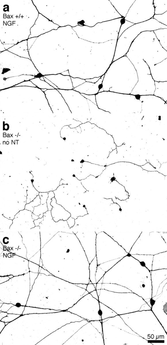
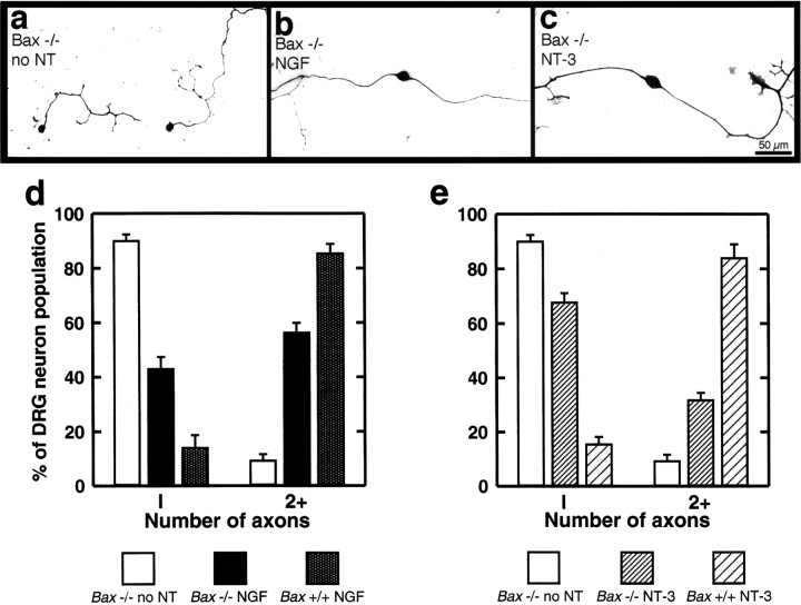
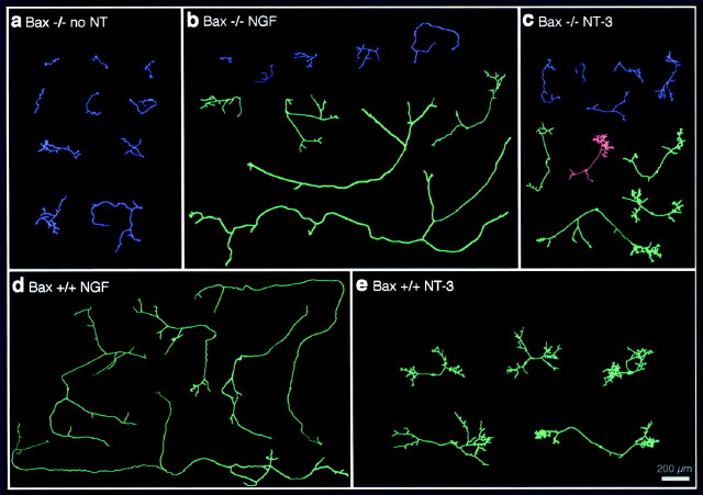
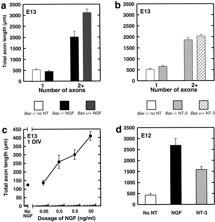
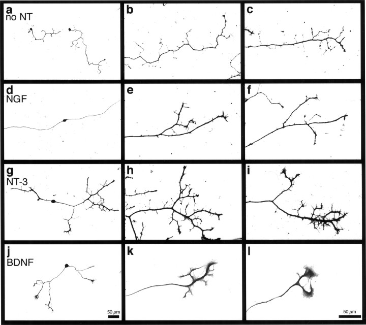
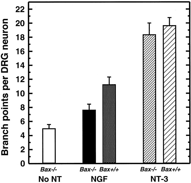
The initial outgrowth of peripheral axons in developing embryos is thought to occur independently of neurotrophins. However, the degree to which peripheral neurons can extend axons and elaborate axonal arborizations in the absence of these molecules has not been studied directly because of exquisite survival requirements for neurotrophins at early developmental stages. We show here that embryonic sensory neurons from BAX-deficient mice survived indefinitely in the absence of neurotrophins, even in highly dissociated cultures, allowing assessment of cell autonomous axon outgrowth. At embryonic day 11 (E11)-E13, stages of rapid axon growth toward targets in vivo, Bax-/- sensory neurons cultured without neurotrophins were almost invariably unipolar and extended only a rudimentary axon. Addition of neurotrophins caused outgrowth of a second axon and a marked, dose-dependent elongation of both processes. Surprisingly, morphological responses to individual neurotrophins differed substantially. Neurotrophin-3 (NT-3) supported striking terminal arborization of subsets of Bax-/- neurons, whereas NGF produced predominantly axon elongation in a different subset. We conclude that axon growth in vitro is neurotrophin dependent from the earliest stages of sensory neuron development. Furthermore, neurotrophins support the appearance of distinct axonal morphologies that characterize different sensory neuron subpopulations.
Figures
Similar articles
-
The cytokine/neurotrophin axis in peripheral axon outgrowth.Eur J Neurosci. 2006 Nov;24(10):2721-30. doi: 10.1111/j.1460-9568.2006.05155.x. Eur J Neurosci. 2006. PMID: 17156198
-
NGF and NT-3 have differing effects on the growth of dorsal root axons in developing mammalian spinal cord.J Neurosci. 1994 Sep;14(9):5187-201. doi: 10.1523/JNEUROSCI.14-09-05187.1994. J Neurosci. 1994. PMID: 8083730 Free PMC article.
-
Absence of sensory neurons before target innervation in brain-derived neurotrophic factor-, neurotrophin 3-, and TrkC-deficient embryonic mice.J Neurosci. 1997 Dec 1;17(23):9113-21. doi: 10.1523/JNEUROSCI.17-23-09113.1997. J Neurosci. 1997. PMID: 9364058 Free PMC article.
-
Role of neurotrophins and trk receptors in the development and maintenance of sensory neurons: an overview.Philos Trans R Soc Lond B Biol Sci. 1996 Mar 29;351(1338):365-73. doi: 10.1098/rstb.1996.0030. Philos Trans R Soc Lond B Biol Sci. 1996. PMID: 8730773 Review.
-
Neurotrophins as in vitro growth cone guidance molecules for embryonic sensory neurons.Cell Tissue Res. 1997 Nov;290(2):285-97. doi: 10.1007/s004410050933. Cell Tissue Res. 1997. PMID: 9321690 Review.
Cited by
-
Mice deficient in brain-derived neurotrophic factor have altered development of gastric vagal sensory innervation.J Comp Neurol. 2010 Aug 1;518(15):2934-51. doi: 10.1002/cne.22372. J Comp Neurol. 2010. PMID: 20533354 Free PMC article.
-
Null mutations of NT-3 and Bax affect trigeminal ganglion cell number but not brainstem barrelette pattern formation.Somatosens Mot Res. 2013 Sep;30(3):114-9. doi: 10.3109/08990220.2013.775118. Epub 2013 Apr 24. Somatosens Mot Res. 2013. PMID: 23614607 Free PMC article.
-
Molecular determinants of the face map development in the trigeminal brainstem.Anat Rec A Discov Mol Cell Evol Biol. 2006 Feb;288(2):121-34. doi: 10.1002/ar.a.20285. Anat Rec A Discov Mol Cell Evol Biol. 2006. PMID: 16432893 Free PMC article. Review.
-
Factors that regulate embryonic gustatory development.BMC Neurosci. 2007 Sep 18;8 Suppl 3(Suppl 3):S4. doi: 10.1186/1471-2202-8-S3-S4. BMC Neurosci. 2007. PMID: 17903280 Free PMC article. Review.
-
Whole-mount imaging of mouse embryo sensory axon projections.J Vis Exp. 2014 Dec 9;(94):52212. doi: 10.3791/52212. J Vis Exp. 2014. PMID: 25549235 Free PMC article.
References
-
- Barde Y-A. Help from within damaged axons. Nature. 1997;385:391–393. - PubMed
-
- Buchman VL, Davies AM. Different neurotrophins are expressed and act in a developmental sequence to promote the survival of embryonic sensory neurons. Development. 1993;118:989–1001. - PubMed
-
- Chen DF, Schneider GE, Martinou J-C, Tonegawa S. Bcl-2 promotes regeneration of severed axons in mammalian CNS. Nature. 1997;385:434–439. - PubMed
-
- Davies AM, Bandtlow C, Heumann R, Korsching S, Rohrer H, Thoenen H. Timing and site of nerve growth factor synthesis in developing skin in relation to innervation and expression of the receptor. Nature. 1987;326:353–358. - PubMed
-
- Davis BM, Fundin BT, Albers KM, Goodness TP, Cronk KM, Rice FL. Overexpression of nerve growth factor in skin causes preferential increases among innervation to specific sensory targets. J Comp Neurol. 1997;387:489–506. - PubMed
Publication types
MeSH terms
Substances
Grants and funding
LinkOut - more resources
Full Text Sources
Molecular Biology Databases
Research Materials