. 1999 Feb 1;19(3):1115-21.
doi: 10.1523/JNEUROSCI.19-03-01115.1999.

Inhibition of light- or glutamate-induced mPer1 expression represses the phase shifts into the mouse circadian locomotor and suprachiasmatic firing rhythms

Affiliations

Inhibition of light- or glutamate-induced mPer1 expression represses the phase shifts into the mouse circadian locomotor and suprachiasmatic firing rhythms

M Akiyama et al. J Neurosci. .

Abstract

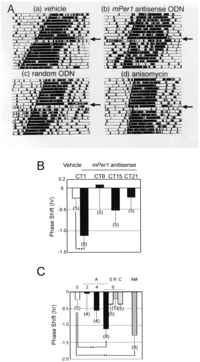
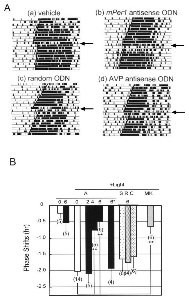
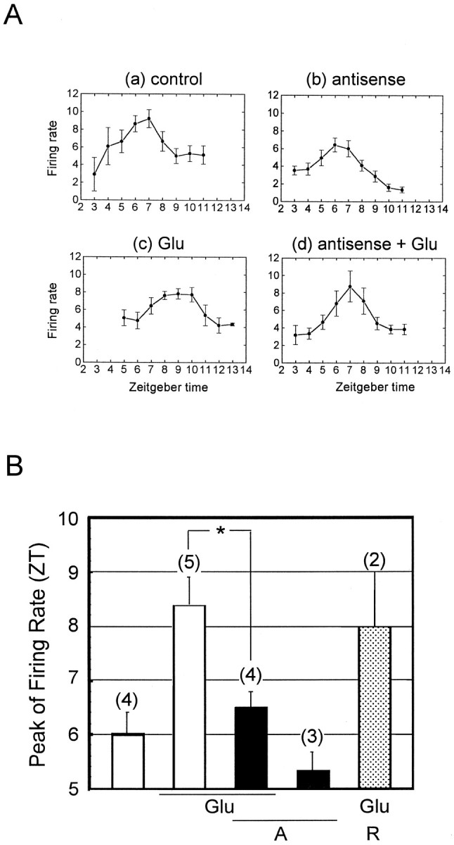
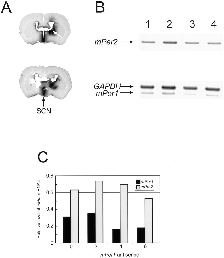
mPer1, a mouse gene, is a homolog of the Drosophila clock gene period and has been shown to be closely associated with the light-induced resetting of a mammalian circadian clock. To investigate whether the rapid induction of mPer1 after light exposure is necessary for light-induced phase shifting, we injected an antisense phosphotioate oligonucleotide (ODN) to mPer1 mRNA into the cerebral ventricle. Light-induced phase delay of locomotor activity at CT16 was significantly inhibited when the mice were pretreated with mPer1 antisense ODN 1 hr before light exposure. mPer1 sense ODN or random ODN treatment had little effect on phase delay induced by light pulses. In addition, glutamate-induced phase delay of suprachiasmatic nucleus (SCN) firing rhythm was attenuated by pretreatment with mPer1 antisense ODN, but not by random ODN. The present results demonstrate that induction of mPer1 mRNA is required for light- or glutamate-induced phase shifting, suggesting that the acute induction of mPer1 mRNA in the SCN after light exposure is involved in light-induced phase shifting of the overt rhythm.


Figures

Fig. 1.
Fig. 1.
Effect of ODN administration at CT1 on the mouse circadian locomotor rhythm. A, Locomotor activity records of vehicle (a), mPer1antisense ODN (b), random ODN (c), and anisomycin (d)-injected mice. Each animal was injected at CT1 (⋄ in the figure) intracerebroventricularly (5 μl; 1 μl min−1) and returned to constant darkness.B, Phase–response curve for mPer1antisense ODN administration at CT1, CT8, CT15, and CT21.Numbers in parentheses indicate the number of experiments. Injection of mPer1 antisense ODN at CT1 induced a significant phase delay (**p < 0.01; Student’s t test). C, Phase shifts of mouse locomotor rhythm by various ODNs or anisomycin injection at CT1.0, Vehicle; A, mPer1antisense ODN; S, sense ODN; R, random ODN; C, AVP antisense ODN. The number in the figure indicates the amount (in nanomoles) of injected ODN.Numbers in parentheses indicate the number of experiments. Injection of mPer1 antisense ODN and anisomycin significantly phase delayed locomotor rhythm (**p < 0.01; Student’s ttest).
Fig. 2.
Fig. 2.
Effect of ODN injection on light-induced phase delay of locomotor activity rhythm. Mice were injected with ODNs at CT15 under the safety light, 1 hr after injection, mice were exposed to light (20 lux) for 15 min and returned to constant darkness.A, Locomotor activity records of vehicle (a), mPer1 antisense (b), random ODN (c), and AVP antisense ODN (d)-injected mouse.B, Light-induced phase shifts in mPer1antisense ODN (A), sense ODN (S), random ODN (R), AVP antisense ODN (C), and MKC-801 (MK)-injected mouse. * indicates that antisense ODN was administered 2 hr after the light pulse. The number in the figure indicates the amount (nanomoles) of injected ODN. 0 indicates the vehicle administration. Numbers in parentheses indicate the number of experiments. Preinjection of mPer1antisense ODN (4 and 6 nmol) and MK-801 significantly reduced light-induced phase shift (**p < 0.01; Student’st test). Injection of mPer1 antisense ODN 2 hr after light exposure did not have any effects.
Fig. 3.
Fig. 3.
Effect of mPer1 antisense ODN on glutamate–induced phase delay of SCN firing rhythm in vitro. A, The average neuronal activity rhythms in the SCN recorded from mice slice on day 2. Each pointindicates the 2 hr means ± SEM of firing rate of single SCN cells from ZT2–14. B, Average phase shifts induced by glutamate and glutamate plus mPer1 antisense ODN. Eachbar indicates the peak of firing rate (mean ± SEM). Numbers in parentheses indicate the number of slices. Preincubation of mPer1 antisense ODN significantly reduced glutamate-induced phase shift (*p < 0.05 vs glutamate alone; Student’st test). Glu, Glutamate;A, mPer1 antisense ODN; R,mPer1 random ODN.
Fig. 4.
Fig. 4.
Effects of mPer1antisense ODN on the mPer1 expression in the SCN.A, Distribution of biotinylated ODN 2 hr after injection into the brain. mPer1 antisense ODN (5′-biotnylated; 6 nmol in 5 ul) was microinjected intracerebroventricularly. Mice were killed 2 hr later, followed by detection of biotinylated ODN. Anarrow on the top slice shows antisense ODN injection site. An arrow on thebottom slice shows the position of SCN. The ODNs were most extensively distributed around the third ventricle including the SCN. B, Inhibition of light induction ofmPer1 transcript in the SCN by in vivo mPer1 antisense ODN treatment. Total RNA was isolated 1.5 hr after light exposure from mPer1 antisense ODN-pretreated mice, and mPer1, mPer2, and GAPDH RNA were amplified by an RT-PCR method. Lane 1, Treated with vehicle; lane 2, treated with 2 nmol of antisense ODN;lane 3, treated with 4 nmol of ODN; lane 4, treated with 6 nmol of antisense ODN. The PCR products ofmPer1, mPer2, and GAPDH gene are indicated by arrows. C, Semiquantitative analysis of RT-PCR products shown in B. The band intensity of RT-PCR products of mPer1 andmPer2 mRNA was measured by one-dimensional analysis software (Eastman Kodak), and their amounts were normalized against GAPDH.

References

    1. Agrawal S, Temsamani J, Tang JY. Pharmacokinetics, biodistribution, and stability of oligodeoxynucleotide phosphorothioates in mice. Proc Natl Acad Sci USA. 1991;88:7595–7599. - PMC - PubMed
    1. Albrecht U, Sun ZS, Eichele G, Lee CC. A differential response of two putative mammalian circadian regulators, mPer1 and mPer2, to light. Cell. 1997;91:1055–1064. - PubMed
    1. Colwell CS, Ralph MR, Menaker M. Do NMDA receptors mediate the effects of light on circadian behavior? Brain Res. 1990;523:117–120. - PubMed
    1. Daan S, Pittendrigh C. A functional analysis of circadian pacemakers in nocturnal rodents II. The variability of phase response curves. J Comp Physiol [A] 1976;106:253–266.
    1. de Vries MJ, Nunes Cardozo B, van der Want J, de Wolf A, Meijer JH. Glutamate immunoreactivity in terminals of the retinohypothalamic tract of the brown Norwegian rat. Brain Res. 1993;612:231–237. - PubMed

Publication types

MeSH terms

LinkOut - more resources