. 1999 Feb;181(3):884-92.
doi: 10.1128/JB.181.3.884-892.1999.

Fibrillar array in the cell wall of a gliding filamentous cyanobacterium

Affiliations

Fibrillar array in the cell wall of a gliding filamentous cyanobacterium

D G Adams et al. J Bacteriol. 1999 Feb.

Abstract

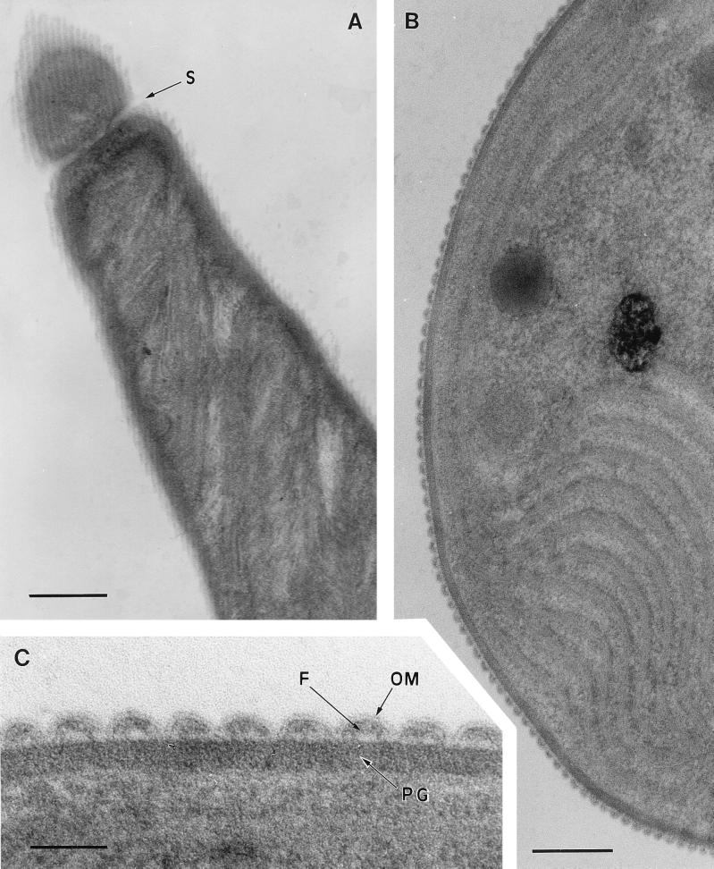
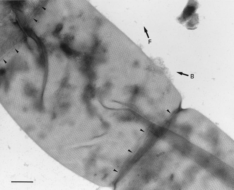
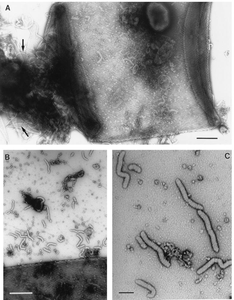
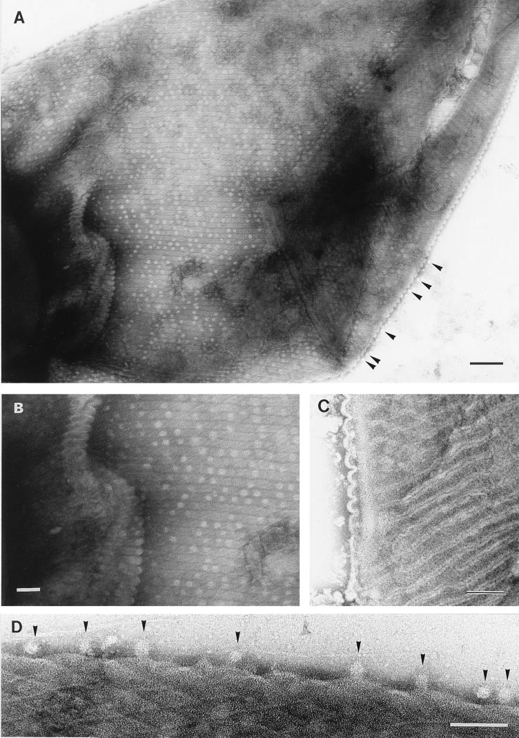
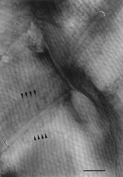
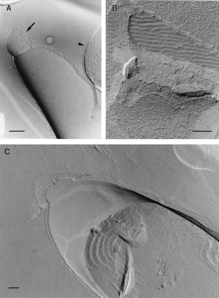
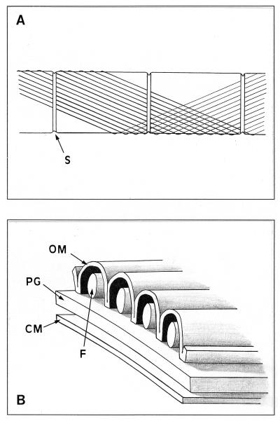
The cell walls of a number of filamentous, gliding cyanobacteria of the genus Oscillatoria were examined by transmission electron microscopy of ultrathin sections, of freeze-etched replicas, and of whole cells crushed between glass slides and negatively stained. All three techniques revealed the presence of a highly ordered array of parallel fibrils, seen in transverse sections to be situated between the peptidoglycan and the outer membrane. Approximately 200 individual fibrils, each 25 to 30 nm in width, form a parallel, helical array that completely surrounds each cyanobacterial filament, running at an angle of 25 to 30 degrees to its long axis. This highly regular arrangement of the fibrillar layer may imply some underlying symmetry responsible for its organization. A possible source of such symmetry would be the peptidoglycan, and some form of interaction between this layer and the fibrils might provide the necessary scaffolding for the fibrillar array. In crushed, negatively stained samples of fresh cells, individual fibrils were seen outside the filament, released from the cell wall. These released fibrils were of the same width as those observed in situ but were in short lengths, mostly of 100 to 200 nm, and were invariably bent, sometimes even into U shapes, implying great flexibility. Negative staining of released fibrils showed no evidence that they were hollow tubes but did give some indication of a substructure, implying that they were composed of many subunits. The function of this fibrillar array is unknown, although its position in the cell wall, as well as the correspondence between the angle of the fibrils with respect to the long axis of the filament and the rotation of the filament during gliding, may imply an involvement in gliding motility.


Figures

FIG. 1
FIG. 1
Transmission electron micrographs of thin sections of Oscillatoria sp. strain FT2 filaments. (A) Longitudinal section. At the top of the figure, beyond a cell septum (S), the section has grazed the surface of the filament, revealing an array of parallel fibrils running at an angle of approximately 25 to 30° to the filament’s long axis. Bar, 400 nm. (B) Part of a transverse section, showing an end view of the fibrillar array in the cell wall. Bar, 200 nm. (C) Enlarged view of part of panel B, showing the double line of the outer membrane (OM) covering the fibrillar array and dipping between adjacent fibrils (F) to contact the electron-dense peptidoglycan layer (PG). Bar, 50 nm.
FIG. 2
FIG. 2
Transmission electron micrograph of part of a filament of Oscillatoria sp. strain FT3. An actively motile sample was crushed between glass slides and negatively stained. The micrograph shows several cells from which the contents have been extruded and the cell wall has been flattened, bringing the fibrils at the front and back of the filament into close contact and thus allowing them to be viewed simultaneously. The fibrils run helically around the entire surface of the filament, producing the observed criss-cross effect because those in the wall in the foreground run in the direction shown by the arrow marked F and those in the wall in the background run in the direction of the arrow marked B. The fibrils appear to cross the cell septa (at the top left and bottom right of the photo), although they are overlayed by a band with a width of approximately 40 nm (arrowheads). Bar, 500 nm.
FIG. 3
FIG. 3
(A and B) Transmission electron micrographs of Oscillatoria sp. strain A2. Actively motile samples were crushed between glass slides and negatively stained. (A) Although an intact fibrillar array covering the cell can be seen, the crushing process has caused damage to the cell wall, leaving some areas (arrows) completely free of fibrils. Some of the fibrils released in this process can be seen in panel B, and some of them show possible evidence of a substructure (C). Bars, 400 nm (A and B) and 100 nm (C).
FIG. 4
FIG. 4
Transmission electron micrographs of Oscillatoria sp. strain FT1. Actively motile samples were crushed between glass slides and negatively stained. (A) The cell walls are covered with regularly arranged circular structures approximately 20 nm in diameter, which are unstained except at the center. These structures are located between the electron-dense lines that separate the fibrils and appear not to be pores, because they can be lost from the surface and seen free, external to the cell (arrowheads here and in panel D). Bar, 200 nm. (B) Shown is an enlarged view of part of panel A, revealing the circular structures that follow the line of each fibril. Bar, 100 nm. (C) The fibrils are separated by an electron-dense line bounded on each side by electron-transparent regions. The former is caused by the accumulation of stain in the cleft in the outer membrane, where it dips between each row of fibrils, and the latter is the unstained membrane itself, on either side of the cleft. These corrugations in the outer membrane can be seen to the left of the micrograph (and are illustrated in Fig. 7). Bar, 100 nm. (D) Enlarged view of part of the edge of the cell from the top left corner of panel A, showing the unstained, 20-nm-diameter circular structures (arrowheads) external to the cell. Bar, 100 nm.
FIG. 5
FIG. 5
Enlarged view of the top left corner of Fig. 2. The cell septum runs from the bottom left to the top right of the photograph. Each fibril is seen as a pale line bounded by two parallel dark lines, and at the point at which they cross the cell septum they appear to pass beneath a narrow band (indicated by the square brackets) that follows the line of the septum. The continuation of three of the fibrils, on either side of the septum, is shown by the arrowheads indicating the dark lines between the fibrils. Bar, 200 nm.
FIG. 6
FIG. 6
Transmission electron micrographs of freeze-etched replicas of Oscillatoria sp. strain A2. (A) Corrugations in the outer membrane, caused by the fibrillar array, can be seen in a transverse view (arrowhead) and in a longitudinal view in the exoplasmic fracture face of the membrane (arrow). (B) Apparent undulations in the fibrillar array are evident. (C) Indentations in the outer membrane, caused by the fibrillar array, can be seen in the exoplasmic fracture face of the membrane, confirming that the fibrillar array itself is external to the cytoplasmic membrane and peptidoglycan. Bars, 400 nm (A) and 200 nm (B and C).
FIG. 7
FIG. 7
Schematic diagrams showing the fibrillar array in the cell wall of Oscillatoria spp. (A) Part of a filament showing three cell septa (S). Each fibril follows a helical path at 25 to 30° to the long axis of the filament. For clarity, only a small number of fibrils and only one turn of the helix are shown here. At the cell septa, the fibrils pass beneath a band that encircles the septum. (B) Cross section of the cell wall, showing the arrangement of the fibrils (F) in relation to the outer membrane (OM), the cytoplasmic membrane (CM), and the peptidoglycan layer (PG).

References

    1. Adams D G. Multicellularity in cyanobacteria. In: Mohan S, Dow C, Cole J A, editors. Prokaryotic structure and function: a new perspective. Cambridge, United Kingdom: Cambridge University Press; 1992. pp. 341–384.
    1. Adams D G. Cyanobacteria. In: Shapiro J A, Dworkin M, editors. Bacteria as multicellular organisms. Oxford, United Kingdom: Oxford University Press; 1997. pp. 109–148.
    1. Guglielmi G, Cohen-Bazire G. Structure et distribution des pores et des perforations de l’enveloppe de peptidoglycane chez quelques cyanobactéries. Protistologica. 1982;18:151–165.
    1. Häder D-P, Hoiczyk E. Gliding motility. In: Melkonian M, editor. Algal cell motility. New York, N.Y: Chapman and Hall; 1992. pp. 1–38.
    1. Halfen L N. Gliding motility in Oscillatoria: ultrastructural and chemical characterization of the fibrillar layer. J Phycol. 1973;9:248–253.

LinkOut - more resources