

Imaging of thrombi and assessment of left atrial appendage function: a prospective study comparing transthoracic and transoesophageal echocardiography
- PMID: 9922358
- PMCID: PMC1728943
- DOI: 10.1136/hrt.81.2.192
Imaging of thrombi and assessment of left atrial appendage function: a prospective study comparing transthoracic and transoesophageal echocardiography
Abstract
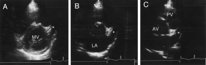
Objective: To compare the value of current transthoracic echocardiographic systems and transoesophageal echocardiography for assessing left atrial appendage function and imaging thrombi.
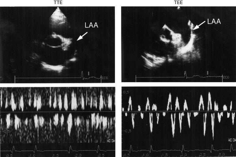
Design: Single blind prospective study. Patients were first investigated by transthoracic echocardiography and thereafter by a second investigator using transoesophageal echocardiography. The feasibility of imaging the left atrial appendage, recording its velocities, and identifying thrombi within the appendage were determined by both methods.
Patients: 117 consecutive patients with a stroke or transient neurological deficit.
Setting: Tertiary cardiac and neurological care centre.
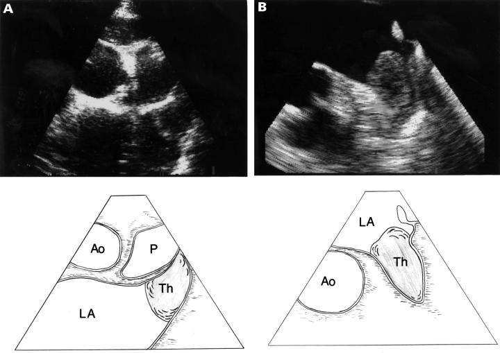
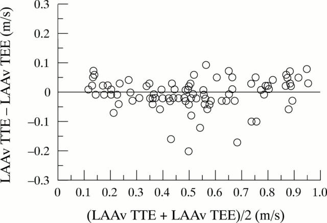
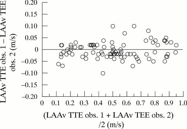
Results: Imaging of the complete appendage was feasible in 75% of the patients by transthoracic echocardiography and in 95% by transoesophageal echocardiography. Both methods were concordant for the detection of thrombi in 10 cases. Transoesophageal echocardiography revealed two additional thrombi. In one of these patients, transthoracic echocardiography was not feasible and in the other the thrombus had been missed by transthoracic examination. In patients with adequate transthoracic echogenicity, the specificity and sensitivity of detecting left atrial appendage thrombi were 100% and 91%, respectively. Recording of left atrial appendage velocities by transthoracic echocardiography was feasible in 69% of cases. None of the patients with a velocity > 0.3 m/s had left atrial appendage thrombi. In the one patient in whom transthoracic echocardiographic evaluation missed a left atrial appendage thrombus, the peak emptying velocity of the left atrial appendage was 0.25 m/s.
Conclusions: A new generation echocardiographic system allows for the transthoracic detection of left atrial appendage thrombi and accurate determination of left atrial appendage function in most patients with a neurological deficit.
Figures
References
Publication types
MeSH terms
LinkOut - more resources
Full Text Sources