

RA/DA cumulative curve analysis of local and diffuse neuroretinal rim area damage in glaucoma patients
- PMID: 9924369
- PMCID: PMC1722672
- DOI: 10.1136/bjo.82.7.769
RA/DA cumulative curve analysis of local and diffuse neuroretinal rim area damage in glaucoma patients
Abstract
Aim: To evaluate the validity of cumulative rim/disc area (RA/DA) curve analysis as a clinical tool for the identification of glaucoma induced optic disc pathology.
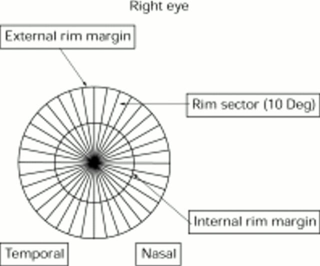
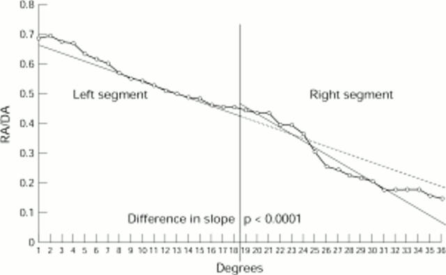
Methods: 71 normal and 83 glaucomatous eyes were evaluated from a series of 154 subjects recruited for this study. For each eye, the cumulative distribution of RA/DA was calculated from 36 equally spaced rim sectors of each optic disc obtained by the automatic evaluation of simultaneous videographics (Image-net X Rev.3/51b). To increase the sensitivity of this analysis in early glaucoma and in normal eyes, these cumulative curves were subsequently divided into two equal segments and the slopes of their respective regression lines compared.
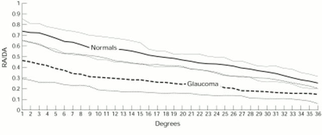
Results: The median RA/DA value obtained from the 36 sectors was significantly different in glaucomatous eyes compared with normals (p < 0.001). Nevertheless, the curves (5th-95th percentile of the cumulative curves distribution) of early glaucomatous eyes fell within the normal range. When the cumulative curve of these marginal cases was then divided into two equal segments, the comparison of the slopes of the regression lines showed a significant difference (p < 0.05) in 100% of early glaucomatous eyes. Furthermore, normal eyes were shown to be true negatives in 93% of the cases in which no significant difference between the two slopes was observed.
Conclusion: Analysis of the RA/DA cumulative curve from 36 sectors of the optic disc was a valid method for the identification of glaucomatous disc pathology; however, a further calculation of the slopes of the two RA/DA regression lines was needed to identify early glaucomatous damage.
Figures
Similar articles
-
Quantitative evaluation of the optic nerve head in early glaucoma.Br J Ophthalmol. 1998 Apr;82(4):352-61. doi: 10.1136/bjo.82.4.352. Br J Ophthalmol. 1998. PMID: 9640180 Free PMC article.
-
The cumulative normalised rim/disc area ratio curve.Graefes Arch Clin Exp Ophthalmol. 1996 Apr;234(4):227-31. doi: 10.1007/BF00430414. Graefes Arch Clin Exp Ophthalmol. 1996. PMID: 8964527
-
[Optic disc measurements with computerized image analysis in experimental chronic glaucoma].Nippon Ganka Gakkai Zasshi. 1989 Aug;93(8):852-8. Nippon Ganka Gakkai Zasshi. 1989. PMID: 2610166 Japanese.
-
Role of Retinal Nerve Fiber Layer Thickness and Optic Disk Measurement by OCT on Early Diagnosis of Glaucoma.Eye Sci. 2015 Mar;30(1):7-12. Eye Sci. 2015. PMID: 26390791
-
Biomorphometry and histomorphometry of the optic disc with special reference to the parapapillary region.Bull Soc Belge Ophtalmol. 1992;244:45-60. Bull Soc Belge Ophtalmol. 1992. PMID: 1297517 Review.
Cited by
-
Long-Term Rate of Optic Disc Rim Loss in Glaucoma Patients Measured From Optic Disc Photographs With a Deep Neural Network.Transl Vis Sci Technol. 2024 Sep 3;13(9):9. doi: 10.1167/tvst.13.9.9. Transl Vis Sci Technol. 2024. PMID: 39235397 Free PMC article.
References
MeSH terms
LinkOut - more resources
Full Text Sources
Medical