. 1999 Jan 15;13(2):176-87.
doi: 10.1101/gad.13.2.176.

Nim1-related kinases coordinate cell cycle progression with the organization of the peripheral cytoskeleton in yeast

Affiliations

Nim1-related kinases coordinate cell cycle progression with the organization of the peripheral cytoskeleton in yeast

Y Barral et al. Genes Dev. .

Abstract

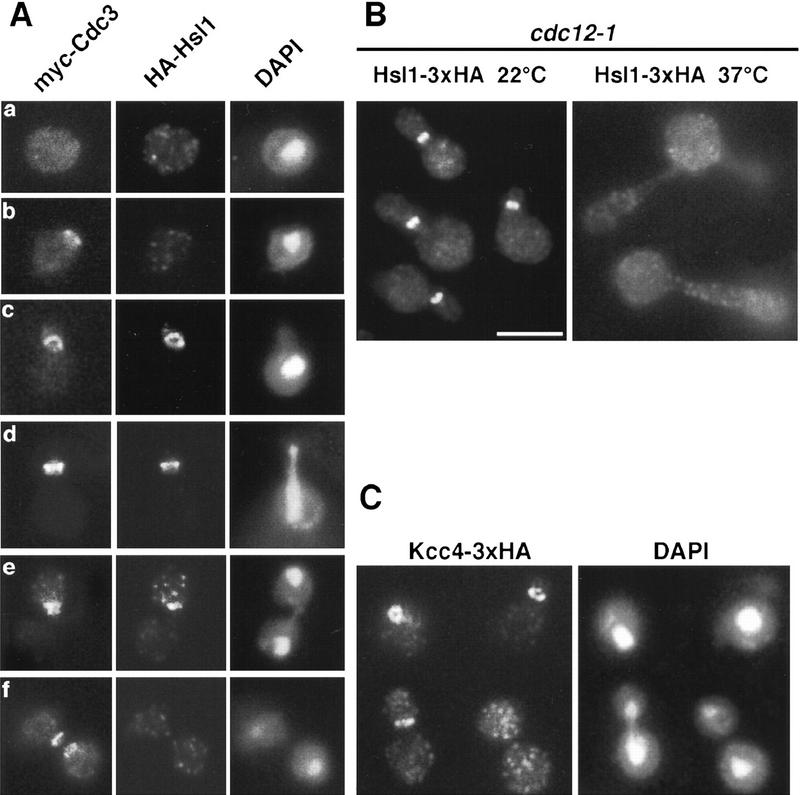
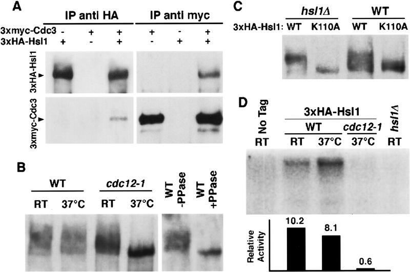
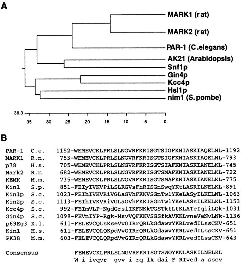
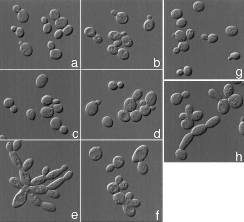
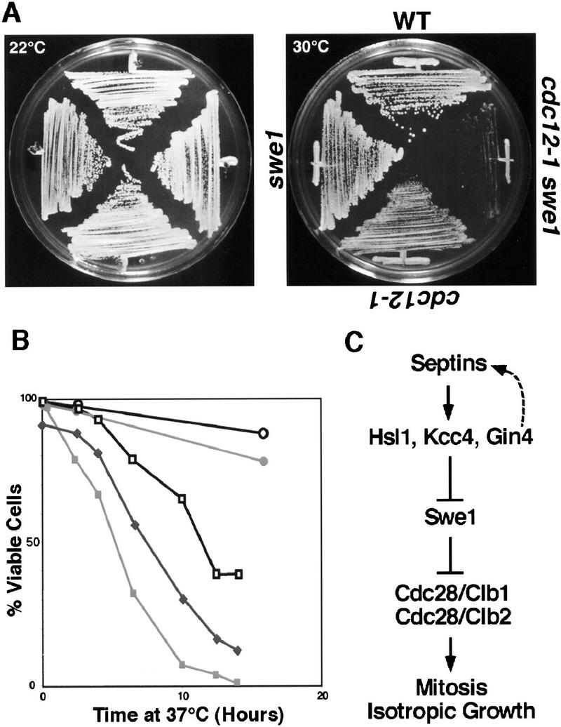
The mechanisms that couple cell cycle progression with the organization of the peripheral cytoskeleton are poorly understood. In Saccharomyces cerevisiae, the Swe1 protein has been shown previously to phosphorylate and inactivate the cyclin-dependent kinase, Cdc28, thereby delaying the onset of mitosis. The nim1-related protein kinase, Hsl1, induces entry into mitosis by negatively regulating Swe1. We have found that Hsl1 physically associates with the septin cytoskeleton in vivo and that Hsl1 kinase activity depends on proper septin function. Genetic analysis indicates that two additional Hsl1-related kinases, Kcc4 and Gin4, act redundantly with Hsl1 to regulate Swe1. Kcc4, like Hsl1 and Gin4, was found to localize to the bud neck in a septin-dependent fashion. Interestingly, hsl1 kcc4 gin4 triple mutants develop a cellular morphology extremely similar to that of septin mutants. Consistent with the idea that Hsl1, Kcc4, and Gin4 link entry into mitosis to proper septin organization, we find that septin mutants incubated at the restrictive temperature trigger a Swe1-dependent mitotic delay that is necessary to maintain cell viability. These results reveal for the first time how cells monitor the organization of their cytoskeleton and demonstrate the existence of a cell cycle checkpoint that responds to defects in the peripheral cytoskeleton. Moreover, Hsl1, Kcc4, and Gin4 have homologs in higher eukaryotes, suggesting that the regulation of Swe1/Wee1 by this class of kinases is highly conserved.


Figures

Figure 1
Figure 1
Hsl1 and Kcc4 localize to the bud neck. (A) A 3xHA–HSL1 3xmyc–CDC3 strain (Y1536) stained with anti-Myc (Myc–Cdc3) and anti-HA (HA–Hsl1) antibodies and DAPI (DNA staining). Cells at different stages of the cell cycle are presented as follows: Unbudded (G1; a,b), small budded (G1/S; c), mitosis (d,e), and cytokinesis (f). Cdc3 forms a ring in unbudded cells (b), a broad ring spanning the neck in budded cells (c), and a double ring in cells that undergo mitosis and cytokinesis (d–f). Hsl1 is not detected in unbudded cells, localizes as a ring at the neck on the bud side in budded cells (c,d,e), and is not evident after cells complete mitosis (f). (Note the ring staining in c). Localization of Hsl1 and Cdc3 is not detected in untagged strains. (B) Localization of Hsl1–3xHA in cdc12-1 cells incubated at either the permissive (left) or restrictive temperature (37°C, right). (C) Indirect immunofluorescence of KCC4–3xHA cells (Y1533) with anti-HA antibodies. Four representative cells from different fields are shown. Kcc4 localizes as a ring in unbudded cells (top left cell) and at the bud side of the neck in budded cells. All pictures are at the same scale. Bar, 5 μm.
Figure 2
Figure 2
Septins and Hsl1 coimmunoprecipitate and Hsl1 autophosphorylation and kinase activity depend on septins. (A) Protein extracts from either 3xmyc–CDC3 3xHA–HSL1 (Y1536), 3xmyc–CDC3 (Y1535), or 3xHA–HSL1 (Y1530) strains were immunoprecipiated with either anti-Myc or anti-HA antibodies. The pellets were separated by SDS-PAGE, and immunoblots were probed with both anti-HA and anti-Myc antibodies. (B) Immunoblot analysis of extracts of 3xHA–HSL1 (Y1530) and 3xHA–HSL1 cdc12-1 (Y1534) cells grown at 24°C and 37°C. High molecular weight isoforms are detected; these are absent in cdc12-1 cells incubated at 37°C and in samples (immunoprecipitates) treated with calf alkaline phosphatase. (C) Plasmids encoding either wild-type HSL1 or a putative kinase-inactive HSL1, hsl1–K110A, were introduced into hsl1Δ (Y1527) and wild-type (Y1521) strains. The phosphorylated isoforms are absent in hsl1–K110A strains and reduced in hsl1–K110A–HSL1 strains. (D) Hsl1 shows little kinase activity when isolated from septin mutants. Anti-HA immunoprecipitates were incubated in the presence of [32P]ATP under kinase reaction conditions. Protein samples were subjected to electrophoresis and exposed for autoradiography. Numbers under each lane correspond to 32P incorporation standardized to the amount of protein as quantified by immunoblotting.
Figure 3
Figure 3
Analysis of Hsl1-related kinases. (A) A dendrogram depicting members of the Nim1/Hsl1 family shows that nim1, Hsl1, Kcc4, and Gin4 are closely related. These alignments were obtained by comparison of the catalytic domains of these enzymes. (B) Sequence analysis reveals that the MARK (microtubule affinity regulatory kinase) kinases, PAR-1, Hsl1, Kcc4, Gin4, and several other kinases of the same family share a 42-amino-acid domain at their noncatalytic, carboxyl terminus. This domain is absent in other members of the family such as Nim1 and Snf1.
Figure 4
Figure 4
hsl1Δ kcc4Δ gin4Δ cells have an elongated cell morphology. (a) Wild-type (Y1521), (b) kcc4Δ (Y1528), (c) gin4Δ (Y1529), (d) hsl1Δ (Y1527), (e) hsl1Δ kcc4Δ gin4Δ (1538), and (f) hsl1Δ kcc4Δ gin4Δ swe1Δ (1537) cells were grown at 22°C and viewed by differential interference contrast microscopy. gin4Δ and hsl1Δ cells exhibit a slight morphological defect, whereas double-mutant (not shown) and triple-mutant cells (e) exhibit an elongated cell morphology; this defect is suppressed by swe1Δ (f). hsl1Δ exacerbates the cdc12-1 morphological defect. cdc12-1 cells grown at the permissive temperature (g) do not have a morphological defect; however, the cdc12–1 hsl1Δ double mutant has elongated buds (h).
Figure 5
Figure 5
Localization of 3xmyc–Cdc3 in wild-type (a) and hsl1Δ kcc4Δ gin4Δ cells (b). Cdc3 localization is aberrant in hsl1Δ kcc4Δ gin4Δ cells but similar to wild type in budded hsl1Δ kcc4Δ gin4Δ swe1Δ cells. Bar, 5 μm.
Figure 6
Figure 6
(A) Septin mutants have an elongated morphology that depends on Swe1. Wild-type (Y1521), cdc12-1 (Y1524), swe1Δ (Y1525), and swe1Δ cdc12-1 cells were grown at 24°C and shifted to 37°C for 3 hr. The temperature-sensitive cdc12-1 cells form elongated buds; this phenotype is suppressed by swe1Δ. Bar, 5 μm. (B,C) Septin mutants exhibit a SWE1-dependent delay in nuclear division. Wild-type (Y1521), cdc12-1 (1524), and swe1Δ cdc12-1 cells were grown at 24°C and arrested with mating pheromone. Cells were diluted into fresh medium lacking pheromone at 37°C, and the percentage of cells that had budded (B) or undergone nuclear division (C) was determined at the indicated times. cdc12-1 cells are delayed prior to nuclear division, which is partially dependent on SWE1. (⋄) wild type; (█) cdc12; (•) cdc12 swe1Δ; (○) cdc 12 CDC28Y–19F.
Figure 7
Figure 7
SWE1 is needed to maintain the viability of septin mutants exposed to the restrictive temperature. (A) The cdc12-1 swe1Δ double mutant has a reduced growth rate at 30°C compared with a cdc12-1 strain. Strains of the indicated phenotypes were incubated at either 22°C or 30°C. (B) Viability assay. Cells of the indicated genotype were incubated at 37°C, samples were taken at various time points, and the percentage of cells that maintained viability was determined by plating at 22°C. (○) wild type; (•) swe1Δ; (□) cdc12; (█) cdc12 swe1Δ; (♦) cdc12 CDC28–Y19F. (C) Model for the septin checkpoint of yeast. Proper assembly of the septin cytoskeleton results in localization and activation of Hsl1, Kcc4, and Gin4. This in turn inactivates Swe1, which thereby allows activation of the Cdc28 kinase. Thus, organization of the septin cytoskeleton is one mechanism by which cells detect that they have assembled both a bud and a proper cleavage apparatus for cytokinesis.

References

    1. Altman R, Kellogg D. Control of mitotic events by Nap1 and the Gin4 kinase. J Cell Biol. 1997;13:119–130. - PMC - PubMed
    1. Baudin A, Ozier-Kalogeropoulos O, Denouel A, Lacroute F, Cullin C. A simple and efficient method for direct gene deletion in S. cerevisiae. Nucleic Acids Res. 1993;21:3329–3330. - PMC - PubMed
    1. Benton B, Tinkelenberg A, Gonzalez I, Cross F. Cla4p, a S. cerevisiae Cdc42p-activated kinase involved in cytokinesis, is activated at mitosis. Mol Cell Biol. 1997;17:5067–5076. - PMC - PubMed
    1. Blacketer MJ, Madaule P, Myers AM. Mutational analysis of morphologic differentiation in S. cerevisiae. Genetics. 1995;140:1259–1275. - PMC - PubMed
    1. Booher RN, Deshaies RJ, Kirschner MW. Properties of S. cerevisiae wee1 and its differential regulation of p34CDC28 in response to G1 and G2 cyclins. EMBO J. 1993;12:3417–3426. - PMC - PubMed

Publication types

MeSH terms