. 1999 Feb 15;515 ( Pt 1)(Pt 1):41-8.
doi: 10.1111/j.1469-7793.1999.041ad.x.

N-linked glycosylation sites determine HERG channel surface membrane expression

Affiliations

N-linked glycosylation sites determine HERG channel surface membrane expression

K Petrecca et al. J Physiol. .

Abstract

1. Long QT syndrome (LQT) is an electrophysiological disorder that can lead to sudden death from cardiac arrhythmias. One form of LQT has been attributed to mutations in the human ether-a-go-go-related gene (HERG) that encodes a voltage-gated cardiac K+ channel. While a recent report indicates that LQT in some patients is associated with a mutation of HERG at a consensus extracellular N-linked glycosylation site (N629), earlier studies failed to identify a role for N-linked glycosylation in the functional expression of voltage-gated K+ channels. In this study we used pharmacological agents and site-directed mutagenesis to assess the contribution of N-linked glycosylation to the surface localization of HERG channels. 2. Tunicamycin, an inhibitor of N-linked glycosylation, blocked normal surface membrane expression of a HERG-green fluorescent protein (GFP) fusion protein (HERGGFP) transiently expressed in human embryonic kidney (HEK 293) cells imaged with confocal microscopy. 3. Immunoblot analysis revealed that N-glycosidase F shifted the molecular mass of HERGGFP, stably expressed in HEK 293 cells, indicating the presence of N-linked carbohydrate moieties. Mutations at each of the two putative extracellular N-linked glycosylation sites (N598Q and N629Q) led to a perinuclear subcellular localization of HERGGFP stably expressed in HEK 293 cells, with no surface membrane expression. Furthermore, patch clamp analysis revealed that there was a virtual absence of HERG current in the N-glycosylation mutants. 4. Taken together, these results strongly suggest that N-linked glycosylation is required for surface membrane expression of HERG. These findings may provide insight into a mechanism responsible for LQT2 due to N-linked glycosylation-related mutations of HERG.


Figures

Figure 1
Figure 1. Effect of tunicamycin on the subcellular localization of transiently expressed HERGGFP in HEK 293 cells
A, subcellular localization of HERGGFP. Native GFP fluorescence overlayed on a transmitted-light image. Arrows indicate HERGGFP localization at surface membranes. B, fluorescence image revealing the subcellular localization of HERGGFP. Arrows indicate HERGGFP localization at surface membranes. C, subcellular localization of HERGGFP in cells treated with 1 μg ml−1 tunicamycin for 28 h. Native GFP fluorescence overlayed on a transmitted-light image. Arrows indicate the perinuclear localization of HERGGFP. D, fluorescence image revealing the subcellular localization of HERGGFP in cells treated with 1 μg ml−1 tunicamycin for 28 h. Representative images of three separate experiments are shown.
Figure 2
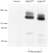
Figure 2. Immunoblot analysis of HERGGFP protein stably expressed in HEK 293 cells
Enriched membrane fractions were prepared as described in Methods. Each lane was loaded with ≈30 μg of protein. An anti-HERG antibody was used to probe for HERGGFP. The control lane shows results with untransfected cells. The HERGGFP lanes show results with cells stably expressing HERGGFP before (-) and after (+) treatment with N-glycosidase F. The data are representative of four separate experiments.
Figure 3
Figure 3. Immunoblot analysis of wild-type and N-linked glycosylation mutants of HERGGFP stably expressed in HEK 293 cells
Enriched membrane fractions were prepared as described in Methods. Each lane was loaded with ≈30 μg of protein. An anti-HERG antibody was used to probe for HERGGFP. The control lane shows results with untransfected cells. Results from protein isolated from cells stably expressing HERGGFP, N598Q-HERGGFP (N598Q), N629Q-HERGGFP (N629Q) or N598Q-N629Q-HERGGFP (N598Q-N629Q) are shown in their respective lanes. The data are representative of four separate experiments.
Figure 4
Figure 4. Whole-cell outward currents in HEK 293 cells stably expressing wild-type and N-linked glycosylation mutants of HERGGFP
A two-step voltage clamp protocol was imposed from a holding potential of -80 mV to assess the presence of HERG current in HEK 293 cells stably transfected with wild-type HERGGFP or HERGGFP mutants. The outward currents were evoked during an initial 4 s depolarizing pulse to potentials between -60 and +50 mV in increments of 10 mV. At the end of the first step the membrane was repolarized back to -60 mV for 2 s before returning to the -80 mV holding potential. Aa, a series of current traces recorded in HEK 293 cells stably transfected with HERGGFP showing an inwardly rectifying delayed rectifier current. Ab, in the presence of E-4031 (1 μM) the HERGGFP-induced delayed rectifier current was completely blocked. The residual transient outward current and background current were similar to those observed in untransfected cells. Ac, current-voltage relationship of untransfected HEK 293 cells (n = 6 cells, ▴) and HEK 293 cells stably transfected with HERGGFP (n = 6, ▪) normalized to the cell capacitance (16.3 ± 1.5 pF; n = 6). B, each panel shows a series of current traces recorded from HEK 293 cells stably transfected with N598Q-HERGGFP, N629Q-HERGGFP or N598Q-N629Q-HERGGFP. HERGGFP current was absent in all three mutants.
Figure 5
Figure 5. Subcellular localization of wild-type and N-linked glycosylation mutants of HERGGFP stably expressed in HEK 293 cells
Cells were immunolabelled with an anti-HERG antibody followed by a FITC-conjugated goat anti-rabbit IgG antibody. Representative immunofluorescence images are overlayed on transmitted-light images. A, surface membrane localization of wild-type HERGGFP. B, perinuclear localization of N598Q-HERGGFP. C, perinuclear localization of N629Q-HERGGFP. D, perinuclear localization of N598Q-N629Q-HERGGFP. Representative images of four separate experiments are shown.

References

    1. Akimoto K, Furutani M, Kasanuki H, Imammura S-I, Furutani Y, Takao A, Monma K, Matsuoka R. Coexistence of missense mutation of HERG and mitochondrial DNA in Japanese long QT family. Circulation. 1996;94:I164.
    1. Benson DW, MacRae CA, Vesely MR, Walsh EP, Seidman JG, Seidman CE, Satler CA. Missense mutation in the pore region of HERG causes familial long QT syndrome. Circulation. 1996;93:1791–1795. - PubMed
    1. Bonifacino JS, Lippincot-Schwartz J. Degradation of proteins within the endoplasmic reticulum. Current Opinion in Cell Biology. 1991;7:592–600. - PubMed
    1. Curran ME, Splawski I, Timothy KW, Vincent GM, Green ED, Keating MT. A molecular basis for cardiac arrhythmia. HERG mutations cause long QT syndrome. Cell. 1995;80:795–803. - PubMed
    1. Dausse E, Berthet M, Denjoy I, Andre-Fouet X, Cruaud C, Bennaceur M, Faure S, Coumel P, Schwartz K, Guicheney P. A mutation in HERG associated with notched T waves in long QT syndrome. Journal of Molecular and Cellular Cardiology. 1996;28:1609–1615. 10.1006/jmcc.1996.0151. - DOI - PubMed

Publication types

MeSH terms

LinkOut - more resources