. 1999 Feb;103(3):331-40.
doi: 10.1172/JCI3858.

Protein 4.1R-deficient mice are viable but have erythroid membrane skeleton abnormalities

Affiliations

Protein 4.1R-deficient mice are viable but have erythroid membrane skeleton abnormalities

Z T Shi et al. J Clin Invest. 1999 Feb.

Abstract

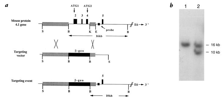
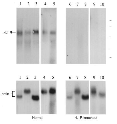
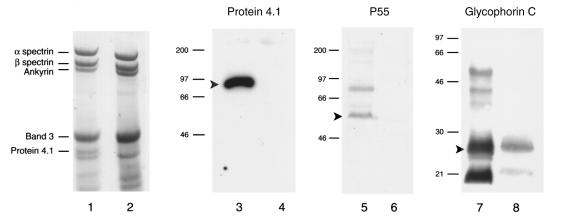
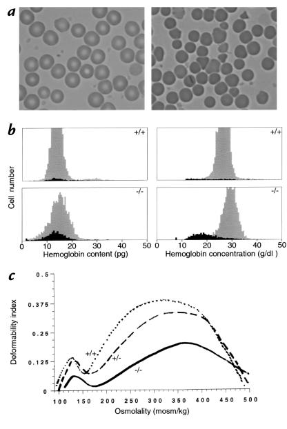
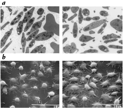
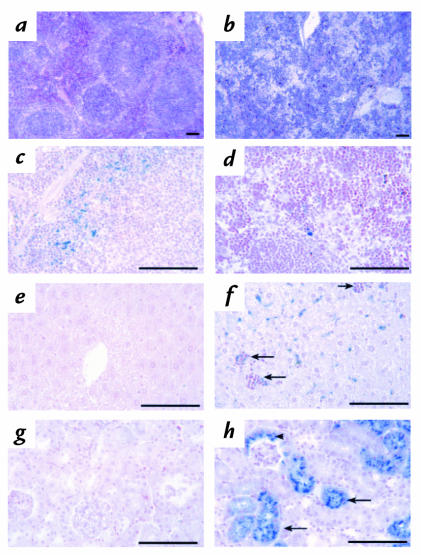
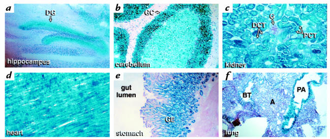
A diverse family of protein 4.1R isoforms is encoded by a complex gene on human chromosome 1. Although the prototypical 80-kDa 4.1R in mature erythrocytes is a key component of the erythroid membrane skeleton that regulates erythrocyte morphology and mechanical stability, little is known about 4.1R function in nucleated cells. Using gene knockout technology, we have generated mice with complete deficiency of all 4.1R protein isoforms. These 4.1R-null mice were viable, with moderate hemolytic anemia but no gross abnormalities. Erythrocytes from these mice exhibited abnormal morphology, lowered membrane stability, and reduced expression of other skeletal proteins including spectrin and ankyrin, suggesting that loss of 4. 1R compromises membrane skeleton assembly in erythroid progenitors. Platelet morphology and function were essentially normal, indicating that 4.1R deficiency may have less impact on other hematopoietic lineages. Nonerythroid 4.1R expression patterns, viewed using histochemical staining for lacZ reporter activity incorporated into the targeted gene, revealed focal expression in specific neurons in the brain and in select cells of other major organs, challenging the view that 4.1R expression is widespread among nonerythroid cells. The 4.1R knockout mice represent a valuable animal model for exploring 4.1R function in nonerythroid cells and for determining pathophysiological sequelae to 4.1R deficiency.


Figures

Figure 1
Figure 1
Targeted disruption of the 4.1R gene. (a) Targeting vector. Top panel depicts a portion of the normal mouse 4.1R gene. Shown are the exon 2–4 region targeted for deletion and the flanking regions upstream (10-kb Sse837I–BamHI fragment) and downstream (2-kb KpnI–EcoRV fragment) that constitute the long arm and short arm of the targeting vector (middle panel). The lower panel represents the structure of the correctly targeted 4.1R gene in which the β-geo cassette has replaced exons 2–4. The targeted 4.1R gene retains the putative upstream promoter and all sequences downstream of exon 5. The intron probe adjacent to exon 5 hybridized to a 16-kb BamHI band in the normal gene and a 10-kb band in the targeted gene. (b) Southern blot showing a successful targeting event in ES cells. In lane 1, control ES cell DNA shows the normal 16-kb BamHI band detected with the intron 4 probe; in lane 2, targeted cell DNA shows the normal 16-kb band and the targeted 10-kb band. ES, embryonic stem.
Figure 2
Figure 2
Analysis of 4.1R mRNA expression by Northern blot analysis. RNA isolated from tissues of a normal control mouse (left) and a homozygous 4.1 knockout mouse (right). Upper panel shows hybridization to a 4.1R cDNA probe representing sequences in exons 18–21, while lower panel shows hybridization to a control actin cDNA probe. Essentially identical results were obtained using a probe that spans exons 2–21. Source of RNA: lanes 1 and 6, heart; lanes 2 and 7, lung; lanes 3 and 8, skeletal muscle; lanes 4 and 9, brain; lanes 5 and 10, spleen. Position of RNA size standards of 9.4, 7.5, 4.4, 2.4, and 1.35 kb is indicated at the top right by the dashed lines.
Figure 3
Figure 3
Distribution of HETE compounds in plaques retrieved from asymptomatic (18 stable plaques) and symptomatic (12 unstable plaques) patients. Each HETE, with concentration expressed on a per-mole-of-lipid-phosphorus basis, taken separately was significantly more abundant in unstable, compared with stable, plaques (P < 0.01).
Figure 4
Figure 4
Erythroid parameters. (a) Red cell morphology in blood from normal (left) and knockout (right) mice. Normal cells at left show the typical biconcave disk morphology, while homozygous 4.1-deficient red cells at right exhibit a more heterogeneous morphology marked by the presence of numerous cell fragments. Red cells from heterozygous mice were morphologically normal. (b) Red cell indices. Cell hemoglobin content and cell hemoglobin concentration histograms of whole blood from normal (upper panels) and knockout (lower panels) mice. The dark areas of the histogram represent reticulocytes, and the gray areas represent mature red cells. Significant numbers of fragmented red cells with decreased hemoglobin content (< 10 pg) can be seen in knockout mice. Cell dehydration is also a feature of knockout mice red cells, as indicated by the shift in hemoglobin concentration from reticulocytes (black area in lower right panel) to mature red cells (gray area in lower right panel). (c). Osmotic gradient deformability profiles of red cells from normal (+/+), heterozygous (+/–), and homozygous (–/–) 4.1R-deficient mice. The maximum value of the deformability index, a direct measure of red cell surface area, is reduced in (+/–) red cells and more dramatically reduced in (–/–) red cells.
Figure 5
Figure 5
Platelet parameters. (a) Transmission electron micrographs of buffy coat fractions of normal (left) and 4.1R-deficient (right) blood. (b) Scanning electron micrographs of platelets adhered to fibrinogen.
Figure 6
Figure 6
Pathological changes in 4.1R–/– tissues. Hematoxylin- and eosin-stained sections of (a) normal and (b) 4.1R–/– spleen sections. Note the effacement of the splenic nodules by the dramatic expansion of the red pulp areas in 4.1R–/– spleen. Prussian blue–stained (c) normal and (d) 4.1R–/– spleen. Iron deposition is decreased in 4.1R–/– spleen red pulp areas due to increased use by rapidly proliferating erythroid precursors. Prussian blue–stained (e) normal and (f) 4.1R–/– liver. Hematopoietic foci (arrows) are frequent and liver iron deposition increased in 4.1R–/– mice, reflecting increased destruction of 4.1-null red cells. Note that no iron deposition is seen in areas of proliferating erythroid precursors, i.e., liver hematopoietic foci (arrows). Prussian blue stains of (g) normal and (h) 4.1R–/– kidney. Note the extensive deposition of iron in 4.1R–/– proximal tubules (arrows) and Bowman's capsule (arrowhead), indicating severe intravascular hemolysis of 4.1R-null red cells. Bar, 100 μM.
Figure 7
Figure 7
Expression of β-galactosidase activity encoded within the β-geo cassette of the targeted 4.1R gene. Frozen sections of the knockout mouse were fixed and stained with the chromogenic substrate 5-bromo-4-chloro-3-indolyl-β-galactosidase, as described in Methods. Expression was observed in the following tissues: (a) hippocampus (b) cerebellum (c) kidney (d) heart (e) stomach, and (f) lung. A, alveoli; BT, bronchiolar tree; DCT, distal convoluted tubule; DG, dentate gyrus; G, glomerulus; GC, granule cells of the cerebellum; GE, gastric epithelium; PA, pulmonary artery; PCT, proximal convoluted tubules.

References

    1. Tchernia G, Mohandas N, Shohet SB. Deficiency of skeletal membrane protein 4.1 in homozygous hereditary elliptocytosis. J Clin Invest. 1981;68:454–460. - PMC - PubMed
    1. Takakuwa Y, Tchernia G, Rossi M, Benabadji M, Mohandas N. Restoration of normal membrane stability to unstable protein 4.1-deficient membranes by incorporation of purified protein 4.1. J Clin Invest. 1986;78:80–85. - PMC - PubMed
    1. Chasis JA, et al. Differentiation-associated switches in protein 4.1 expression. Synthesis of multiple structural isoforms during normal human erythropoiesis. J Clin Invest. 1993;91:329–338. - PMC - PubMed
    1. Anderson RA, Correas I, Mazzucco C, Castle JD, Marchesi VT. Tissue-specific analogues of erythrocyte protein 4.1 retain functional domains. J Cell Biochem. 1988;37:269–284. - PubMed
    1. Granger BL, Lazarides E. Membrane skeletal protein 4.1 of avian erythrocytes is composed of multiple variants that exhibit tissue-specific expression. Cell. 1984;37:595–607. - PubMed

Publication types