. 1999 Feb;103(3):393-9.
doi: 10.1172/JCI4890.

Cell-surface protein disulfide isomerase catalyzes transnitrosation and regulates intracellular transfer of nitric oxide

Affiliations

Cell-surface protein disulfide isomerase catalyzes transnitrosation and regulates intracellular transfer of nitric oxide

A Zai et al. J Clin Invest. 1999 Feb.

Abstract

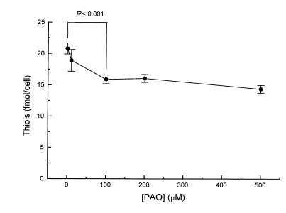
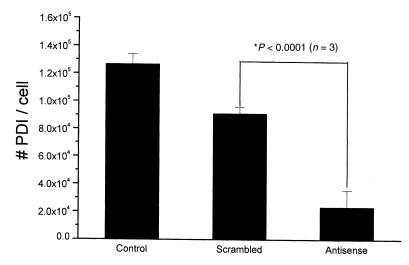
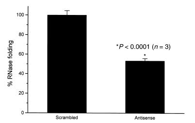
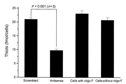
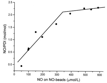
Since thiols can undergo nitrosation and the cell membrane is rich in thiol-containing proteins, we considered the possibility that membrane surface thiols may regulate cellular entry of NO. Recently, protein disulfide isomerase (PDI), a protein that catalyzes thio-disulfide exchange reactions, has been found on the cell-surface membrane. We hypothesized that cell-surface PDI reacts with NO, catalyzes S-nitrosation reactions, and facilitates NO transfer from the extracellular to intracellular compartment. We observed that PDI catalyzes the S-nitrosothiol-dependent oxidation of the heme group of myoglobin (15-fold increase in the rate of oxidation compared with control), and that NO reduces the activity of PDI by 73.1 +/- 21.8% (P < 0.005). To assess the role of PDI in the cellular action of NO, we inhibited human erythroleukemia (HEL) cell-surface PDI expression using an antisense phosphorothioate oligodeoxynucleotide directed against PDI mRNA. This oligodeoxynucleotide decreased cell-surface PDI content by 74.1 +/- 9.3% and PDI folding activity by 46.6 +/- 3.5% compared with untreated or "scrambled" phosphorothioate oligodeoxynucleotide-treated cells (P < 0.0001). This decrease in cell-surface PDI was associated with a significant decrease in cyclic guanosine monophosphate (cGMP) generation after S-nitrosothiol exposure (65.4 +/- 26.7% reduction compared with control; P < 0.05), with no effect on cyclic adenosine monophosphate (cAMP) generation after prostaglandin E1 exposure. These data demonstrate that the cellular entry of NO involves a transnitrosation mechanism catalyzed by cell-surface PDI. These observations suggest a unique mechanism by which extracellular NO gains access to the intracellular environment.


Figures

Figure 1
Figure 1
Quantification of cell-surface thiols. HEL cells were incubated for 10 min with increasing concentrations of PAO, and the remaining free surface thiols were measured by the DTNB assay. We observed that each cell contains 20 ± 0.9 fmol of thiols in which 23.6 ± 3.8% are vicinal dithiols. DTNB, 5,5′-dithiobis(2-nitrobenzoic acid); HEL, human erythroleukemia; PAO, phenylarsine oxide.
Figure 2
Figure 2
Suppression of cell-surface PDI expression. HEL cells were treated with an IgG polyclonal antibody against PDI and a secondary FITC-labeled anti–rabbit antibody. We observed under confocal microscopy a significant decrease in cell-surface PDI expression when HEL cells were preincubated with antisense phosphorothioate against PDI mRNA for 24 h (a) compared with the scrambled control (b). PDI, protein disulfide isomerase.
Figure 3
Figure 3
Effects of antisense phosphorothioate oligodeoxynucleotides on PDI content. The total PDI content of HEL cells was assessed by immunoblotting analysis. We observe a significant decrease in total PDI (lowest band, ∼57 kDa) in the HEL cells preincubated with antisense phosphorothioates against PDI mRNA compared with controls (scrambled phosphorothioates).
Figure 4
Figure 4
Suppression of cell-surface PDI expression. Antisense phosphorothioate blockade of PDI translation suppressed the expression of cell-surface PDI by 74.1 ± 9.2% compared with scrambled phosphorothioates, as detected by fluorescent-labeled anti-PDI antibody.
Figure 5
Figure 5
Reduction in cell-surface PDI folding activity with antisense treatment. Refolding of GS-RNase T1 was performed at 25°C in PBS (pH 7.4). RNase folding activity was measured as extent of folding at 45 min; 100% activity corresponds to the maximal folding by control HEL cells. GS-RNase T1 (6 μM final concentration) was added to incubation mixtures containing 10,000 cells/μl. In antisense-treated cells, folding activity decreased by 46.6 ± 3.5% compared with control. GS-RNase, glutathionyl-RNase T1.
Figure 6
Figure 6
PDI maintains the redox state of cell-surface thiols. HEL cells were treated with an antisense phosphorothioate against PDI mRNA. We observed a 53.6 ± 2.3% decrease in surface thiols, as detected by the DTNB assay.
Figure 7
Figure 7
PDI interacts with NO. PDI (1 μM) was incubated for 10 min with an increasing concentration of SNO-4B. Using the Saville assay, we found that NO binds to PDI in a dose-dependent manner with an NO/PDI ratio of 2:1 at saturation. SNO-4B, S-nitroso-glutathionyl–Sepharose 4B.
Figure 8
Figure 8
PDI, NO, and soluble guanylyl cyclase activation. To determine the role of PDI in the transfer of NO into HEL cells, we measure intracellular cyclic GMP levels. We observed a 65.4 ± 26.7% decrease in cyclic GMP levels in antisense phosphorothioate–treated cells incubated with 10 μM SNO-4B for 10 min compared with scrambled phosphorothioate–treated controls.

References

    1. Lambert N, Freedman R. The latency of rat liver microsomal protein disulfide-isomerase. Biochem J. 1985;228:635–645. - PMC - PubMed
    1. Koch G. Reticuloplasmins: a novel group of proteins in the endoplasmic reticulum. J Cell Sci. 1985;87:491–492. - PubMed
    1. Creighton TE, Hillson D, Freedman R. Catalysis by protein-disulphide isomerase of the unfolding and refolding of proteins with disulphide bonds. J Mol Biol. 1980;142:43–62. - PubMed
    1. Gilbert H. Molecular and cellular aspects of thiol-disulfide exchange. Adv Enzymol Relat Areas Mol Biol. 1990;63:69–172. - PubMed
    1. Gilbert H. Protein disulfide isomerase and assisted protein folding. J Biol Chem. 1997;272:29399–29402. - PubMed

Publication types

MeSH terms