. 1999 Feb 1;189(3):521-30.
doi: 10.1084/jem.189.3.521.

Type I interferons keep activated T cells alive

Affiliations

Type I interferons keep activated T cells alive

P Marrack et al. J Exp Med. .

Abstract

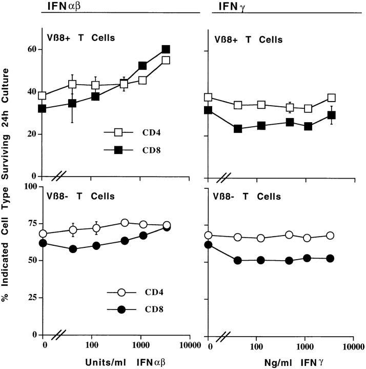
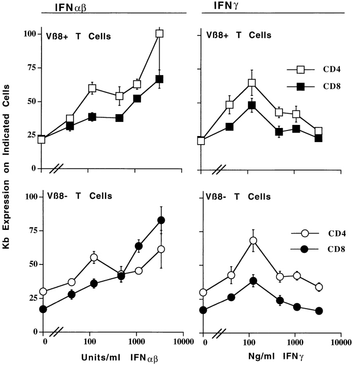
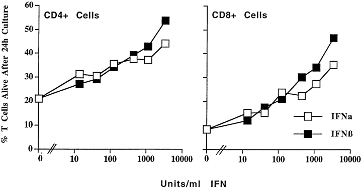
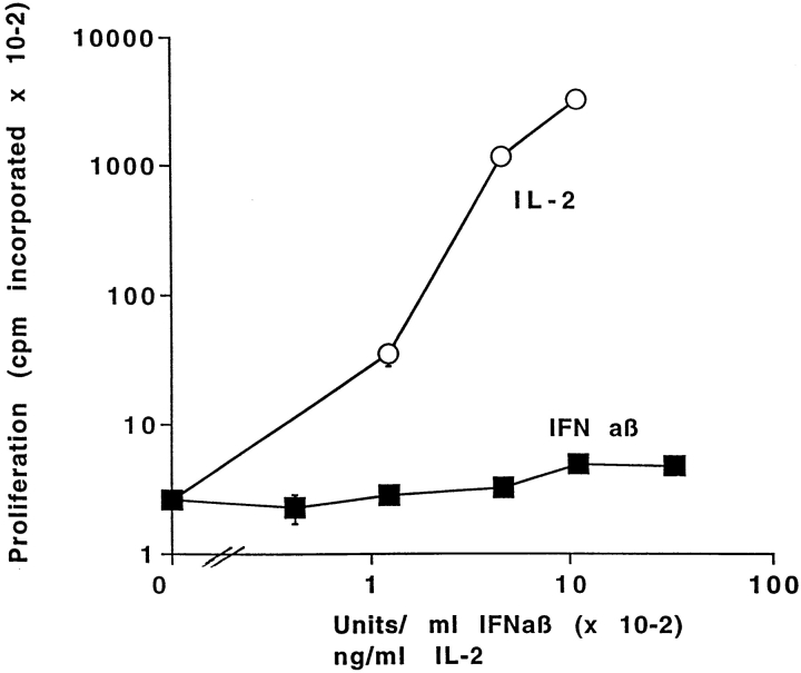
Antigen injection into animals causes antigen-specific T cells to become activated and, rapidly thereafter, die. This antigen-induced death is inhibited by inflammation. To find out how inflammation has this effect, various cytokines were tested for their ability to interfere with the rapid death of activated T cells. T cells were activated in vivo, isolated, and cultured with the test reagents. Two groups of cytokines were active, members of the interleukin 2 family and the interferons (IFNs) alpha and beta. This activity of IFN-alpha/beta has not been described previously. It was due to direct effects of the IFNs on the T cells and was not mediated by induction of a second cytokine such as interleukin 15. IFN-gamma did not slow the death of activated T cells, and therefore the activity of IFN-alpha/beta was not mediated only by activation of Stat 1, a protein that is affected by both classes of IFN. IFN-alpha/beta did not raise the levels of Bcl-2 or Bcl-XL in T cells. Therefore, their activity was distinct from that of members of the interleukin 2 family or CD28 engagement. Since IFN-alpha/beta are very efficiently generated in response to viral and bacterial infections, these molecules may be among the signals that the immune system uses to prevent activated T cell death during infections.


Figures

Figure 1
Figure 1
IFN-α/β prevent the death of activated T cells. T cells were isolated from the lymph nodes of mice given 150 μg SEB 2 d previously. The cells were cultured for 24 h in the absence or presence of 3,333 U/ml IFN-α/β. The cells were then stained with FL-anti-CD4, biotinylated anti-Vβ8x, or anti-Vβ6 followed by PE-labeled streptavidin and Cy-labeled anti-CD8. These cells were then identified as live or dead based on their light scatter properties (top). Separate wells of cells cultured under identical conditions were stained with FL-anti-Vβ6 or anti-Vβ8. These cells were then permeabilized with saponin and incubated with propidium iodide to measure their DNA content. Light scatter gates were set for the permeabilized cells and propidium iodide staining was assessed as shown (bottom). Results shown are typical of duplicate cultures. Values shown on the figure are the percentage of the indicated cells that were alive as defined by the method used.
Figure 2
Figure 2
IFN-α/β prevent the death of activated T cells. IFN-γ does not. T cells were isolated from the lymph nodes of control B10 mice, or from B10 mice that had been injected with 150 μg SEB 2 d previously. The cells were cultured for 20 h in the presence of the indicated concentrations of IFN-α/β or IFN-γ. The percentages of live Vβ8+ or Vβ8 cells in the cultures were determined by flow cytometry as described in the legend to Fig. 1, using light scatter properties as an indicator of survival. Results shown are the mean ±SE of triplicate cultures.
Figure 3
Figure 3
Activated and resting T cells bear receptors for IFN-α/β and IFN-γ. Mice were primed and T cells were cultured as described in the legend to Fig. 2. 20 h after the start of culture, the cells were isolated and stained for Vβ8, Kb, and CD4 or CD8 as described in Materials and Methods. The results shown are the mean ± SE of triplicate cultures of staining with anti-Kb antibody in arbitrary units based on staining intensities.
Figure 4
Figure 4
Both IFN-α and IFN-β affect the survival of activated T cells. T cells were activated and purified as described in the legend to Fig. 1. The cells were cultured for 24 h in the presence of various concentrations of IFN-α or IFN-β and the percentages of live activated (Vβ8+) T cells were determined by flow cytometry. Results shown are the mean ± SE of triplicate cultures.
Figure 5
Figure 5
IFN-α/β do not stimulate the proliferation of activated T cells. Lymph node T cells were purified from B10 mice primed 2 d previously with 150 μg/mouse SEB. The cells were cultured for 3 d in the presence of the indicated concentrations of IFN-α/β or IL-2 and then pulsed with [3H]TdR as described in Materials and Methods Results shown are the mean ± geometric SE of triplicate cultures. Similar results were obtained from cells tested after 2 d of culture.

References

    1. Webb S, Morris C, Sprent J. Extrathymic tolerance of mature T cells: clonal elimination as a consequence of immunity. Cell. 1990;63:1249–1256. - PubMed
    1. Kawabe Y, Ochi A. Programmed cell death and extrathymic reduction of Vβ8+ CD4+ cells in mice tolerant to Staphylococcus aureusenterotoxin B. Nature. 1991;349:245–248. - PubMed
    1. McCormack JE, Callahan JE, Kappler J, Marrack P. Profound deletion of mature T cells in vivo by chronic exposure to exogenous superantigen. J Immunol. 1993;150:3785–3792. - PubMed
    1. Kearney ER, Pape KA, Loh DY, Jenkins MK. Visualization of peptide-specific immunity and peripheral tolerance induction in vivo. Immunity. 1994;1:327–339. - PubMed
    1. Duke RC. IL-2 addiction: withdrawal of growth factor activates a suicide program in dependent T cells. Lymphokine Res. 1986;5:289–299. - PubMed

Publication types

MeSH terms