Comparative Study
. 1999 Feb 1;189(3):531-40.
doi: 10.1084/jem.189.3.531.

Immune responses to Ro60 and its peptides in mice. I. The nature of the immunogen and endogenous autoantigen determine the specificities of the induced autoantibodies

Affiliations
Comparative Study

Immune responses to Ro60 and its peptides in mice. I. The nature of the immunogen and endogenous autoantigen determine the specificities of the induced autoantibodies

U S Deshmukh et al. J Exp Med. .

Abstract

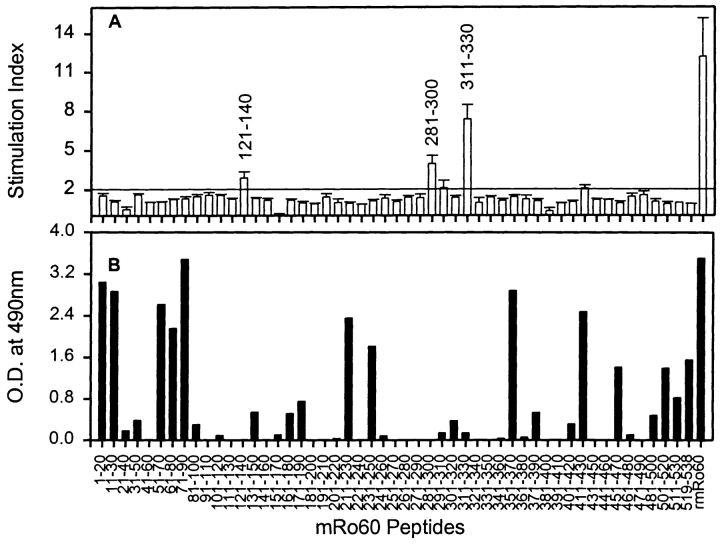
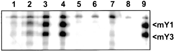
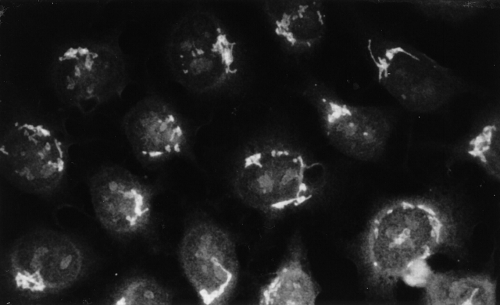
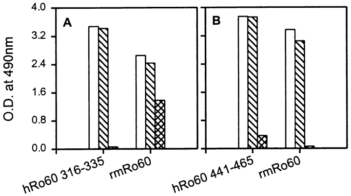
Anti-Ro60 autoantibodies are found in a variety of autoimmune disorders including systemic lupus erythematosus (SLE), Sjögren's syndrome, primary biliary cirrhosis, and active hepatitis. They are the most prevalent autoantibodies in normal individuals and in asymptomatic mothers of infants afflicted with neonatal lupus. In the present study, immune responses to recombinant human Ro60 (rhRo60) and recombinant mouse Ro60 (rmRo60) and selected Ro60 peptides in non-SLE-prone mice were investigated. Multiple T and B cell epitopes were identified in Ro60. Immunizations with either xenogeneic or autologous Ro60 induced autoantibodies to a diverse group of autoantigens. In addition to La and Ro52, proteins in the small nuclear ribonucleoprotein (snRNP) particles such as SmA, SmB, SmD, and 70-kD U1-RNP were unexpectedly identified as targeted antigens. In the studies involving synthetic Ro60 peptides, both human and mouse Ro60316-335 peptides, which differ in three amino acids, were found to contain dominant cross-reactive T cell determinants. Immunizations with these peptides induced autoantibodies to Ro60, La, SmD, and 70-kD U1-RNP without autoantibodies to Ro52, SmA, or SmB. With human Ro60316-335 as the immunogen, additional autoantibodies reactive with the Golgi complex were found. In contrast to the immunodominance of both human and mouse Ro60316-335 peptides, the T cell determinant in human Ro60441-465 was dominant, whereas that in the mouse peptide was cryptic. Immunization with human Ro60441-465 induced primarily anti-peptide Abs. Mouse Ro60441-465 failed to induce an antibody response. These results show that both the nature of the immunogen and the immunogenicity of the related endogenous antigen are important in determining the specificities of the autoantibodies generated. They have significant implications for proposed mechanisms on the generation of complex patterns of autoantibodies to a diverse group of autoantigens in SLE patients.


Figures

Figure 1
Figure 1
Immune response to rhRo60 in SJL/J (circles), A/J (squares), and BALB/c (triangles) mice immunized with rhRo60. T cell responses were studied in LNC proliferative assays (A) and antibody responses were determined by ELISA (B). The LNC proliferative responses are expressed as mean triplicate ΔCPM. In A, results are shown for mice immunized with rhRo60 (open symbols) and with CFA (filled symbols). (B) The ELISA results are expressed as mean duplicate OD490nm and are shown for day 14 (•••••) and day 30 (−) pooled sera. Sera from control mice immunized with CFA, gave OD490nm < 0.1 at a serum dilution of 1:100.
Figure 2
Figure 2
Delineation of T cell determinants on hRo60 in mice of different haplotypes. In vitro LNC proliferative responses were recalled, with synthetic peptides at 20 μM concentration. Results are expressed as SI, a ratio of the mean triplicate CPM obtained with peptide and the mean triplicate cpm without peptide. (A) Multiple determinants recognized by SJL/J T cells are present in Ro60. Results are shown as mean triplicate SI ± SEM from three independent experiments. Peptides giving an SI > 2 are marked with an asterisk (*). For comparison, the response to rhRo60 at a concentration of 3 μg/ml is shown. (B) A summary of T cell determinants on hRo60, seen by SJL/J, A/J, and BALB/c mice. Amino acid sequences for peptides giving a SI > 2 are shown.
Figure 3
Figure 3
Mapping of B cell determinants on hRo60. Sera from, SJL/J, A/J, and BALB/c strains of mice (four/group), immunized with rhRo60, were pooled and reactivity to overlapping synthetic peptides of hRo60 was determined in ELISA. Sera were used at a dilution of 1:500. Results are expressed as mean duplicate OD490nm and are shown for sera obtained on day 14 (▪) and day 30 (□) after immunization. Sera obtained from mice immunized with CFA alone gave a OD490nm < 0.05 at 1:500 dilution.
Figure 4
Figure 4
T cell proliferative responses to rmRo60 in mice of different haplotypes: SJL/J (• rmRo60, ○ CFA), A/J (▪ rmRo60, □ CFA), and BALB/c (▾ rmRo60, ▿ CFA). Results are expressed as mean triplicate ΔCPM. Data for SJL/J strain of mice are represented on the left y-axis. Data for A/J and BALB/c mice are plotted on the right y-axis.
Figure 5
Figure 5
Mapping of T and B cell determinants on mRo60. (A) In vitro T cell proliferative responses were recalled with synthetic peptides of mRo60, at 20 μM concentration. Results are expressed as mean of SI ± SEM from three independent experiments. A mean SI > 2.0 was considered positive. For comparison, the response to rmRo60 at a concentration of 10 μg/ml is included. (B) Reactivity of pooled sera (day 67) at a dilution of 1:500, was checked with synthetic peptides of mRo60 in ELISA. Results are expressed as mean duplicate OD490nm.
Figure 6
Figure 6
Induction of ANAs following immunization with Ro60 antigens. Sera obtained on day 67, postimmunization, were pooled and used at a dilution of 1:200 to stain methanol-fixed HeLa cells (detected by indirect immunofluorescence). a, b, and c represent sera from mice immunized with rmRo60, rhRo60, and CFA, respectively.
Figure 7
Figure 7
Intermolecular determinant spreading of antibody responses in SJL/J mice immunized with Ro60 antigens. Reactivities of sera with different ribonucleoproteins are shown in slot blots. Each lane represents a serum sample at a dilution of 1:250.
Figure 8
Figure 8
Immunoprecipitation of mYRNAs associated with mRo60. WEHI 7.1 cells were labeled with 32P, and RNA species were immunoprecipitated and visualized by autoradiography. Lanes 1–4 represent pooled sera on days 24, 37, 60, and 90, respectively, from SJL/J mice immunized with hRo60316–335. Lanes 5–7, represent pooled sera on days 37, 60, and 90, respectively, from mice immunized with CFA. Lane 8 is control, without any serum, and lane 9 is a human anti-Ro60 reference serum from the Center for Disease Control.
Figure 9
Figure 9
Reactivity of pooled immune sera with WEHI 7.1 cell extracts in Western blot. Each lane was loaded with protein equivalent to 2.5 × 10⁶ cells. Pooled sera were used at a dilution of 1:100. Lane 1, human anti-La reference serum from CDC; lanes 2–4, day 37, 60, and 90 sera from mice immunized with peptide hRo60316–335; lanes 5–7, days 37, 60, and 90 sera from mice immunized with CFA.
Figure 10
Figure 10
Intermolecular determinant spreading of antibody responses in SJL/J mice immunized with hRo60 peptides. Reactivity of sera from mice immunized with peptides hRo60316–335, hRo60441–465, and ZP3 peptide JS7A, with different ribonucleoproteins was tested in slot blots. Each lane represents an individual serum, at a dilution of 1:250.
Figure 11
Figure 11
Anti-Golgi staining patterns of antibodies generated by hRo60316–335 immunization. HeLa cells grown on coverslips and fixed in methanol were used as substrate. Results are shown for pooled sera (day 60) used at a dilution of 1:200.
Figure 12
Figure 12
Intramolecular diversification of antibody responses against mRo60 following immunization with synthetic peptides. Pooled sera from mice immunized either with hRo60316–335 peptide (A), or hRo60441–465 peptide (B) were absorbed with their respective immunogens and peptide JS7A. Reactivity of unabsorbed and absorbed sera with peptides and rmRo60 was determined in ELISA. Results are expressed as mean duplicate OD490nm. Open bars denote unabsorbed pooled sera. Cross bars denote pooled sera, absorbed with peptide JS7A. The hatched bars denote pooled sera absorbed with hRo60316–335 in (A) and with hRo60441–461 in (B).
Figure 13
Figure 13
Recall of in vitro LNC proliferative responses by rmRo60 (open bars) and peptides, mRo60316–335 (cross bars) and mRo60441–465 (hatched bars), in mice immunized with rmRo60. Results are expressed as mean triplicate SI. And an SI > 2.0 was considered positive.
Figure 14
Figure 14
Cross priming between human and mouse peptides. SJL/J mice were immunized with peptides: hRo60316–335 (A), mRo60316–335 (B), hRo60441–465 (C), and mRo60441–465 (D). 2 wk later, proliferative responses were recalled with peptides hRo60316–335 (•), mRo60316–335 (○), hRo60 (▴), mRo60 (▵), hRo60441–465 (▪), and mRo60441–465 (□). Results are expressed as mean triplicate SI. y-axis scales for A, B and C, D are different. Peptide concentrations are in μM and those for Ro60 in μg/ml as shown in the x-axis.
Figure 15
Figure 15
Induction of antibody diversification in mice immunized with peptide mRo60316–335. (A) Reactivity of day 30 sera from four mice with different ribonucleoproteins was tested in slot blots. All sera were tested at 1:250 dilution. (B) Reactivity of pooled sera from SJL/J mice immunized with hRo60316–335 peptide (lane 1), JS7A (lane 2), and CFA (lane 3). (C) Pooled sera from mice immunized with mRo60316–335 were absorbed with the immunogen (hatched bars) and control peptide JS7A (cross bars). The reactivities of the unabsorbed (open bars) and the absorbed sera were determined in ELISA with either mRo60316–335 (left) or mRo60 (right) as the target antigen. The sera absorbed with the immunogen did not react with the immunogen mRo60316–335 while the reactivity with mRo60 remained. Results are expressed as mean duplicate OD490nm.

References

    1. Tan EM, Cohen AS, Fries JF, Masi AT, McShane DJ, Rothfield NF, Schaller JG, Talal N, Winchester RJ. The 1982 revised criteria for the classification of systemic lupus erythematosus (SLE) Arthritis Rheum. 1982;25:1271–1277. - PubMed
    1. Tan EM. Antinuclear antibodies: diagnostic markers for autoimmune disease and probes for cell biopsy. Adv Immunol. 1989;44:93–151. - PubMed
    1. Reichlin, M., and J.B. Harley. 1997. Antinuclear antibodies: an overview. In Dubois' Lupus Erythematosus. D.J. Wallace and B.H. Hahn, editors. Williams and Wilkins, Baltimore, MD. 397–405.
    1. Harley JB, Scofield RH, Reichlin M. Anti-Ro in Sjögren's syndrome and systemic lupus erythematosus. Rheum Dis Clin North Am. 1992;18:337–358. - PubMed
    1. Topfer F, Gordon T, McCluskey J. Intra- and intermolecular spreading of autoimmunity involving the nuclear self-antigens LA (SS-B) and Ro (SS-A) Proc Natl Acad Sci USA. 1995;92:875–879. - PMC - PubMed

Publication types

MeSH terms