. 1999 Feb 2;96(3):893-8.
doi: 10.1073/pnas.96.3.893.

Single-molecule fluorescence spectroscopy of enzyme conformational dynamics and cleavage mechanism

Affiliations

Single-molecule fluorescence spectroscopy of enzyme conformational dynamics and cleavage mechanism

T Ha et al. Proc Natl Acad Sci U S A. .

Abstract

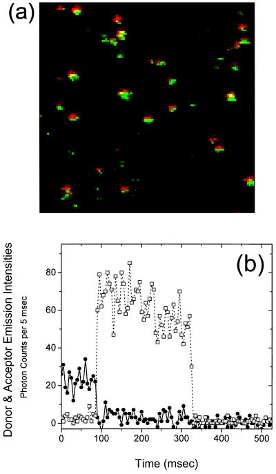
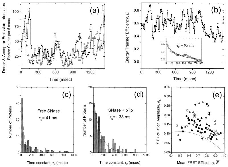
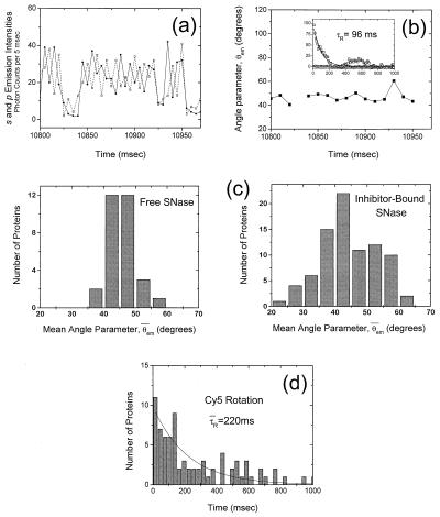
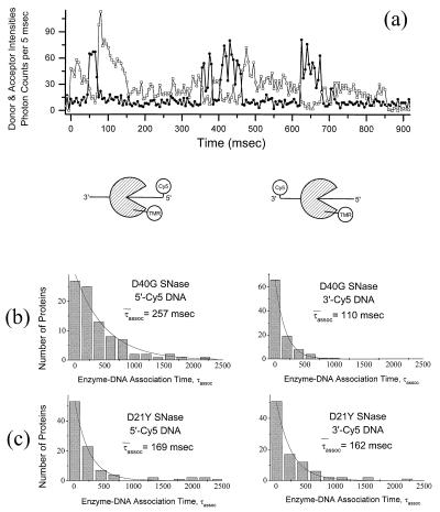
Fluorescence resonance energy transfer and fluorescence polarization anisotropy are used to investigate single molecules of the enzyme staphylococcal nuclease. Intramolecular fluorescence resonance energy transfer and fluorescence polarization anisotropy measurements of fluorescently labeled staphylococcal nuclease molecules reveal distinct patterns of fluctuations that may be attributed to protein conformational dynamics on the millisecond time scale. Intermolecular fluorescence resonance energy transfer measurements provide information about the dynamic interactions of staphylococcal nuclease with single substrate molecules. The experimental methods demonstrated here should prove generally useful in studies of protein folding and enzyme catalysis at single-molecule resolution.


Figures

Figure 1
Figure 1
(a) Dual-color composite image of doubly labeled SNase molecules immobilized by means of histidine tag in buffer A at 23°C. The 514-nm excitation laser spot (Ar+ light, 15 μW) was scanned from top to bottom and left to right. Each spot represents a single protein. Donor emission is colored green and acceptor emission is colored red. (b) Emission time trace of a single double-labeled SNase molecule. The resolution of the measurement is 5 msec. The instantaneous donor and acceptor emission intensities, Id and Ia (squares and circles, respectively), are related to the energy transfer efficiency by the expression E(t) = [1 + γId/Ia]−1, where γ is a correction factor determined to be 0.8. The SNase molecule shown here displays a very high degree of energy transfer (high acceptor intensity, low donor intensity) from 0 to 80 msec. At 80 msec, photodestruction of the acceptor occurs and donor emission simultaneously increases. The inverse correlation is direct evidence for spFRET.
Figure 2
Figure 2
(a) Emission time trace of a doubly labeled TMR/Cy5 SNase molecule immobilized on glass in buffer A with an oxygen scavenging system (donor emission is dotted, acceptor emission is solid). A median filter was applied to reduce the noise caused by triplet state-induced fluorophore blinking. There are large and gradual fluctuations in Id and Ia that occur over tens of milliseconds. (b) FRET efficiency time trace calculated from a according to E(t) = [1 + γId/Ia]−1. The inset shows the autocorrelation of E(t) together with an exponential fit. τE is the characteristic time scale of the E(t) fluctuations displayed in this trace. (c) Histogram of E(t) fluctuation time constants τE for 100 doubly labeled SNase molecules. The values range from 10 msec to 1 sec, with the average being 41 msec. (d) Histogram of E(t) fluctuation time constants τE for doubly labeled SNase in the presence of active-site inhibitor pTp (50 mM pTp, Kd = 100 nM). These time constants are considerably larger (average = 133 msec) than those measured for free SNase. (e) Scatter plot of E(t) fluctuation amplitudes aE vs. mean energy transfer efficiencies Ē for free SNase (squares) and pTp-bound SNase (circles). The scatter in Ē may reflect the nonspecific nature of Cy5 labeling. The solid line represents the maximum possible contribution of dipole fluctuations of Cy5 to aE for free SNase. Only the molecules that display large E(t) fluctuations (>70% of the total) are shown. The others are concentrated around the bottom right corner (not shown) and are likely caused by Cy5 labeling at a site close to Cys28.
Figure 3
Figure 3
(a) A typical smFPA time trace of Cys28 TMR-labeled SNase immobilized by means of histidine tag in buffer A. The two orthogonally polarized emissions, Is (dotted) and Ip (solid), are displayed as a function of time. Because correlated emissions correspond to a fixed fluorophore dipole and anticorrelated emissions correspond to a rapidly rotating fluorophore dipole, the SNase-conjugated TMR molecule shown here is rotating rapidly (much faster than the data integration time of 5 msec). (b) The angle parameter θem(t) calculated from a. The value of θem(t) is close to 45° when the fluorophore is rotating rapidly with little restriction (14). The break in graph represents a dark-state transition. In the inset is the autocorrelation of the angle parameter (in circles); there are clearly no significant temporal fluctuations in TMR rotation on the millisecond time scale. Also in the inset is the angle parameter autocorrelation for pTp-bound Cys28 TMR-labeled SNase (squares) together with an exponential fit. Here the rotational fluctuations are substantial, with a characteristic time constant τR of 96 msec. (c) Histograms of θ̄ for immobilized TMR-labeled SNase molecules with and without inhibitor pTp. The distribution is narrowly centered at 45° for uninhibited SNase, indicative of free and rapid rotation of the attached TMR fluorophore. The TMR of inhibitor-bound SNase, on the other hand, displays hindered and fluctuating rotational behavior, indicated by the broader mean angle parameter histogram; (d) Histogram of rotational fluctuation time constants τR for Cy5-labeled SNase. Only those molecules that showed single-step photobleaching were included to screen out multiply Cy5-labeled cases. The average value of τR is 220 msec, considerably longer than the majority of E(t) fluctuation time constants (average τE = 41 msec, Fig. 2c) .
Figure 4
Figure 4
(a) Emission time trace of a single TMR-labeled wild-type SNase molecule interacting with single Cy5-labeled 40-nt ssDNA molecule(s) (donor emission in squares, acceptor emission in circles). The donor signal decreases every time the acceptor signal increases (signifying the initiation of energy transfer) and vice versa (signifying the termination of energy transfer); this is direct evidence for spFRET. (b) Histograms of τassoc for D40G SNase with 5′-Cy5-ssDNA and 3′-Cy5-ssDNA. The average duration of the FRET signals measured from interactions between donor-labeled SNase and 5′-end acceptor-labeled substrate are longer (average = 257 msec) than those measured with 3′-end acceptor-labeled substrate (average = 110 msec). This difference is consistent with a SNase cleavage mechanism in which the 5′ cleavage product is released more slowly than the 3′ cleavage product, or the enzyme catalyzes cleavage processively in the 3′ to 5′ direction. (c) Histograms of DNA-SNase association times, τassoc, for cleavage-impaired D21Y SNase with 5′-Cy5-ssDNA (left; average τassoc = 169 msec) and 3′-Cy5-ssDNA (right; average τassoc = 162 msec). This control experiment demonstrates that fluorophore photophysics and statistical aberrations do not account for the differences in association times observed when using 5′- and 3′-end-labeled substrates with the cleavage-competent D40G SNase mutant.

References

    1. Funatsu T, Harada Y, Tokunaga M, Saito K, Yanagida T. Nature (London) 1995;374:555–559. - PubMed
    1. Sase I, Miyata H, Corrie J E, Craik J S, Kinosita K. Biophys J. 1995;69:323–328. - PMC - PubMed
    1. Vale R D, Funatsu T, Pierce D W, Romberg L, Harada Y, Yanagida T. Nature (London) 1996;380:451–453. - PMC - PubMed
    1. Schmidt T, Schutz G J, Baumgartner W, Gruber H J, Schindler H. Proc Natl Acad Sci USA. 1996;93:2926–2929. - PMC - PubMed
    1. Sase I, Miyata H, Ishiwata S, Kinosita K. Proc Natl Acad Sci USA. 1997;94:5646–5650. - PMC - PubMed

Publication types

Substances

LinkOut - more resources