. 1999 Feb 2;96(3):915-20.
doi: 10.1073/pnas.96.3.915.

Expression cloning of LDLB, a gene essential for normal Golgi function and assembly of the ldlCp complex

Affiliations

Expression cloning of LDLB, a gene essential for normal Golgi function and assembly of the ldlCp complex

J E Chatterton et al. Proc Natl Acad Sci U S A. .

Abstract

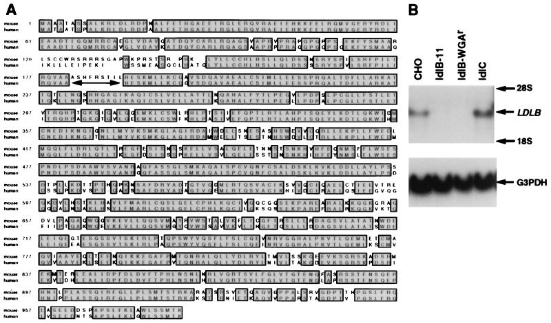
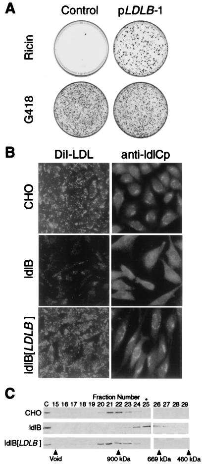
The Chinese hamster ovary (CHO) cell mutants ldlC and ldlB, which exhibit almost identical phenotypes, define two genes required for multiple steps in the normal medial and trans Golgi-associated processing of glycoconjugates. The LDLC gene encodes ldlCp, an approximately 80-kDa protein, which in wild-type, but not ldlB, cells associates reversibly with the cytoplasmic surface of the Golgi apparatus. Here, we have used a retrovirus-based expression cloning system to clone a murine cDNA, LDLB, that corrects the pleiotropic mutant phenotypes of ldlB cells. The corresponding mRNA was not detected in ldlB mutants. LDLB encodes an approximately 110-kDa protein, ldlBp, which lacks homology to known proteins and contains no common structural motifs. Database searches identified short segments of homology to sequences from Drosophila melanogaster, Arabidopsis thaliana, and Caenorhabditis elegans, and the essentially full-length homologous human sequence (82% identity); however, as was the case for ldlCp, no homologue was identified in Saccharomyces cerevisiae. We have found that in wild-type cell cytosols, ldlCp is a component of an approximately 950-kDa "ldlCp complex," which is smaller, approximately 700 kDa, in ldlB cytosols. Normal assembly of this complex is ldlBp-dependent and may be required for Golgi association of ldlCp and for the normal activities of multiple luminal Golgi processes.


Figures

Figure 1
Figure 1
Sequences of the murine and human LDLB cDNAs and LDLB expression in wild-type and mutant CHO cells. (A) Predicted amino acid sequence of murine ldlBp and alignment with a human ldlBp sequence assembled from the EST database (see Materials and Methods). The predicted amino acid sequence is numbered assuming that the first potential initiator codon is utilized. The human and mouse genes were aligned utilizing clustx. Identical amino acids are shaded and boxed. The double-headed, horizontal arrow indicates a gap in the human ldlBp sequence for which no information was available in the EST database. (B) Northern blot analysis (see Materials and Methods) of the LDLB mRNA in wild-type and mutant CHO cells by using LDLB (Upper) or G3PDH (Bottom) cDNA probes.
Figure 2
Figure 2
LDLB cDNA corrects the mutant phenotypes of ldlB cells. (A) Ricin sensitivity. ldlB cells were transfected with either pLDLB-1 or pcDNA3.1 (control). Two days later, the cells were replated and selected with ricin (medium E) or G418 (medium I, transfection efficiency control). Twelve days later, the dishes were fixed and stained with crystal violet. (B) LDLR activity and Golgi localization of ldlCp. Left, wild-type CHO, ldlB, and ldlB[LDLB] cells were grown on glass cover slips in medium F, and LDLR activity, as measured by uptake of fluorescent DiI-LDL (1 μg protein per ml, 1 hr), was observed by using a ×40 objective lens as described (see Materials and Methods). Right, Wild-type CHO, ldlB, and ldlB[LDLB] cells were grown on glass cover slips in medium B and immunostained with a polyclonal anti-ldlCp antibody followed by FITC-labeled goat anti-rabbit IgG and observed by using a ×63 objective lens as described (see Materials and Methods). Golgi localization of ldlCp in CHO and ldlB[LDLB] cells was confirmed by double staining with anti-β-COP antibody (not shown). (C) Gel chromatographic analysis of ldlCp. Cytosol was prepared from wild-type CHO, ldlB, and ldlB[LDLB] cells and analyzed by Superose 6 chromatography as described (see Materials and Methods). Samples were subjected to SDS/PAGE and immunoblotting with affinity-purified polyclonal anti-ldlCp antibody. Fractions containing the ≈80-kDa ldlCp band, as well as unfractionated, diluted (1:10) cytosol (C) are shown. The column was calibrated with Mr standards ranging from 65 to 900 kDa, including apoferritin (460 kDa), thyroglobulin (669 kDa), and IgM (900 kDa) (vertical arrows). The peak fraction of β-COP-containing coatomer, determined by immunoblotting with a monoclonal antibody against β-COP (not shown), is indicated by ∗.

References

    1. Pryer N K, Wuestehube L J, Schekman R. Annu Rev Biochem. 1992;61:471–516. - PubMed
    1. Rothman J E. Nature (London) 1994;372:55–63. - PubMed
    1. Kornfeld R, Kornfeld S. Annu Rev Biochem. 1985;54:631–664. - PubMed
    1. Hirschberg C B, Snider M D. Annu Rev Biochem. 1987;56:63–87. - PubMed
    1. Kingsley D M, Krieger M. Proc Natl Acad Sci USA. 1984;81:5454–5458. - PMC - PubMed

Publication types

MeSH terms

Associated data