. 1999 Feb 2;96(3):1024-9.
doi: 10.1073/pnas.96.3.1024.

T cell antigen receptor-mediated activation of the Ras/mitogen-activated protein kinase pathway controls interleukin 4 receptor function and type-2 helper T cell differentiation

Affiliations

T cell antigen receptor-mediated activation of the Ras/mitogen-activated protein kinase pathway controls interleukin 4 receptor function and type-2 helper T cell differentiation

M Yamashita et al. Proc Natl Acad Sci U S A. .

Abstract

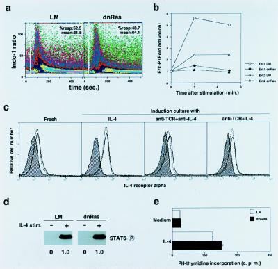
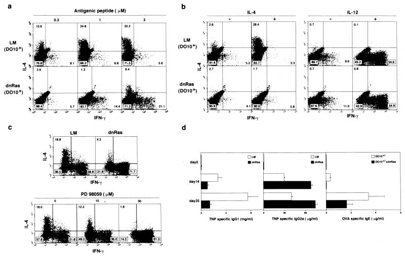
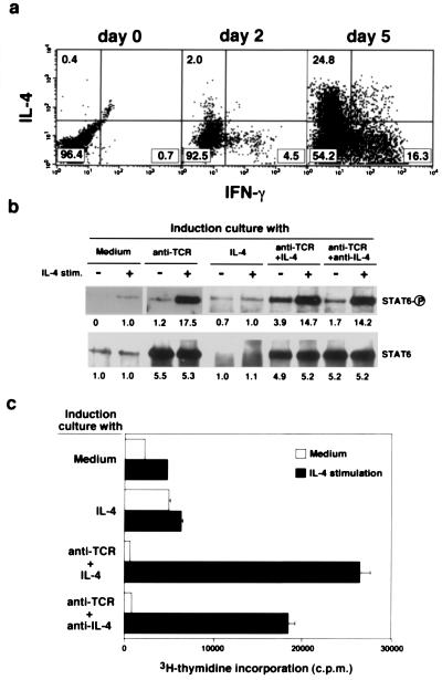
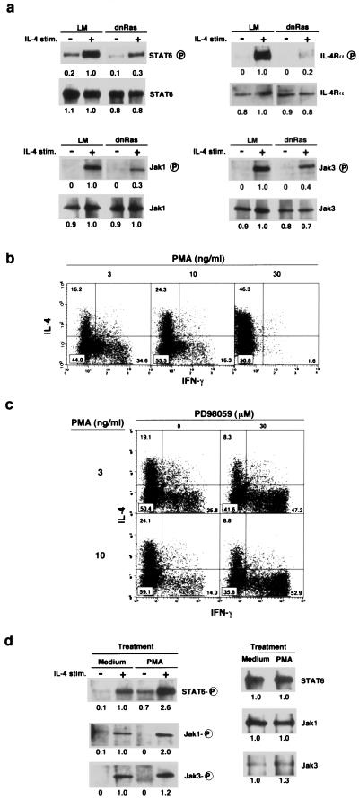
The central role of type-2 helper T (Th2) cells in the development of allergic responses and immune responses against helminthic parasites is well documented. The differentiation of Th2 cells from naive T cells requires both the recognition of antigen by T cell antigen receptors (TCR) and the activation of downstream signal-transduction molecules of the interleukin 4 receptor (IL-4R) pathway, including Jak1, Jak3, and STAT6. Little is known, however, about how these two distinct pathways cooperate with each other to induce Th2 cells. Here, we use a T cell-specific H-Ras-dominant-negative transgenic mouse to show that TCR-mediated activation of the Ras/mitogen-activated protein kinase pathway alters IL-4R function and is required for Th2 cell differentiation. The enhancement of IL-4R signaling seems to be a consequence of both direct "crosstalk" with the TCR signaling pathway and increased protein expression of downstream signaling molecules of the IL-4R pathway. Therefore, successful Th2 differentiation depends on the effectiveness of the TCR-mediated activation of the Ras/mitogen-activated protein kinase pathway in modifying the IL-4R-mediated signaling pathway.


Figures

Figure 1
Figure 1
Signal transduction through TCR and IL-4R in naive CD4 T cells from dnRas Tg mice. (a) Intracellular free-calcium ion levels after TCR crosslinking (at white gap) were measured by flow cytometric analysis of Indo-1-labeled naive CD4 T cells of dnRas Tg mice and littermate (LM) controls. The mean ratio of violet to blue fluorescence of Indo-1 is plotted versus time after stimulation. Shown are data obtained by gating electronically on CD4 T cells. The percentages of responding cells and mean percentages are also shown. (b) Phosphorylation status of MAPK (Erk1 and Erk2) was examined 2 min and 5 min after TCR crosslinking by using a phospho-MAPK detection kit. Shown is the relative increase (fold) in phosphorylation of Erk1 and Erk2 with respect to unstimulated control cells (time 0). Densitometric measurement was used for the quantification; three independent experiments were done, and similar results were obtained. (c) Cell-surface expression of IL-4Rα chain on naive CD4 T cells from Tg LM mice (dotted line) and from dnRas Tg mice (solid line) was determined after a 2-day induction culture with indicated stimulants. Background staining is shown (hatched areas). (d) Tyrosine phosphorylation of STAT6 in response to IL-4 in naive CD4 T cells from dnRas Tg mice. Freshly prepared CD44low T cells were stimulated with IL-4 (100 units/ml) for 5 min, and the tyrosine phosphorylation status of STAT6 was assessed by immunoblotting with anti-phosphotyrosine mAb (RC20). Arbitrary densitometric units are shown under each band. (e) Normal proliferative response to IL-4 (100 units/ml for 40 h) by naive CD4 T cells from dnRas Tg mice.
Figure 2
Figure 2
Requirement for activation of Ras/MAPK pathway in Th2 cell differentiation in vitro and in vivo. (a) Naive CD4 T cells from dnRas × DO10 double-Tg mice were stimulated with antigenic peptide (OVA; 323-339) and irradiated BALB/c (H-2d) antigen-presenting cells for 5 days, and intracellular production of IFN-γ and IL-4 was detected. (b) Naive T cells were stimulated with either a minimal dose (0.3 μM) of antigenic peptide and exogenous IL-4 (100 units/ml; Left) or with 1 μM antigenic peptide in the presence of anti-IL-4 mAb and exogenous IL-12 (0.1 unit/ml; Right). (c) Th1/Th2 cell differentiation of naive T cells from dnRas Tg mice with (B6 × BALB/c)F1 background and the effect of a specific inhibitor of MEK (MAPKK), PD 98059. Naive CD4 T cells from normal and dnRas Tg mice were stimulated with immobilized anti-TCR in the presence of IL-2 (30 units/ml) and IL-4 (100 units/ml) for 2 days and then cultured in the medium with the same concentrations of IL-2 and IL-4 for another 3 days. The cultured cells were subjected to intracellular staining with anti-IL-4 and anti-IFN-γ. (d) Effect of overexpression of dnRas on helper T cell differentiation in vivo. Tg LM mice and dnRas Tg mice were primed on day 0 and boosted on day 21; 2 weeks after each immunization, serum concentrations of antigen-specific antibodies were determined by ELISA. Mice with B6 backgrounds were immunized with TNP-keyhole limpet hemocyanin (100 μg per mouse) in complete Freund’s adjuvant, and serum concentrations of TNP-specific IgG1 and IgG2a were measured (Left and Center). DO10 × dnRas Tg control (D10+/−) and DO10 × dnRas double-Tg (D10+/− × dnRas) mice were immunized with OVA (100 μg per mouse) in alum, and serum concentrations of OVA-specific IgE were measured (Right). Bars depict mean values of four animals with standard deviations expressed as error bars.
Figure 3
Figure 3
Improvement of IL-4R-mediated signal transduction after TCR stimulation. (a) Anti-TCR-induced Th1/Th2 cell differentiation on day 2 and day 5. Anti-TCR-stimulated naive CD4 T cells from (B6 × BALB/c)F1 mice were harvested on day 0, day 2, and day 5. The percentages of cells in each area are shown. (b) Naive CD4 T cells from (B6 × BALB/c)F1 mice were cultured for 2 days with medium, immobilized anti-TCR mAb, IL-4 alone, immobilized anti-TCR mAb and IL-4, or immobilized anti-TCR and anti-IL-4 mAb (induction culture). The percentages of recovered live cells in these cultures were 92%, 123%, 89%, 120%, and 135%. The induced cells were cultured without IL-4 or anti-IL-4 for another 8 h at 37°C, and then the cells were stimulated with IL-4 (100 units/ml) for 5 min. The phosphorylation status of STAT6 and the amount of STAT6 protein were assessed by immunoprecipitation with anti-STAT6 mAb and, after immunoblotting, with anti-phosphotyrosine or anti-STAT6 mAb. The equivalent of 20 million cells was loaded in each lane. (c) In vitro cultured cells like those in b were stimulated with IL-4 (100 units/ml) for 40 h, and [3H]thymidine incorporation (over the last 16 h) was measured.
Figure 4
Figure 4
IL-4-induced phosphorylation on STAT6 and Jak1 in naive T cells was regulated by the Ras/MAPK signaling pathway. (a) Naive CD4 T cells from dnRas Tg mice were cultured for 2 days with immobilized anti-TCR mAb and IL-4 and cultured without IL-4 for 8 h. Then, IL-4-induced phosphorylation on STAT6, IL-4Rα, Jak1, and Jak3 was assessed by immunoprecipitation with specific mAb for each protein and, after immunoblotting, with anti-phosphotyrosine mAb. Arbitrary densitometric units are shown under each band. The amount of protein was determined also by reblotting the same membrane with specific mAbs. (b) Phorbol 12-myristate 13-acetate (PMA) dose-dependent increase in the generation of Th2 cells in vitro. Naive T cells from (B6 × BALB/c)F1 mice were treated with indicated doses of PMA for 2 days in the presence of ionomycin (300 nM), IL-2 (30 units/ml), and IL-4 (100 units/ml). (c) Effect of PD98059 on the PMA-induced Th1/Th2 cell differentiation. (d) Naive CD4 T cells were treated with PMA (30 ng/ml) for 4 h, and IL-4-induced phosphorylation on STAT6, Jak1, and Jak3 was assessed by the same method used in a. The amount of each protein existing in the cells with or without PMA treatment was also determined (Right).

References

    1. Mosmann T R, Cherwinski H, Bond M W, Giedlin M A, Coffman R L. J Immunol. 1986;136:2348–2357. - PubMed
    1. Seder R A, Paul W E. Annu Rev Immunol. 1994;12:635–673. - PubMed
    1. Abbas A K, Murphy K M, Sher A. Nature (London) 1996;383:787–793. - PubMed
    1. O’Garra A. Immunity. 1998;8:275–283. - PubMed
    1. Seder R A, Gazzinelli R, Sher A, Paul W E. Proc Natl Acad Sci USA. 1993;90:10188–10192. - PMC - PubMed

Publication types

MeSH terms