. 1999 Feb 2;96(3):1100-5.
doi: 10.1073/pnas.96.3.1100.

Neuroligin 1 is a postsynaptic cell-adhesion molecule of excitatory synapses

Affiliations

Neuroligin 1 is a postsynaptic cell-adhesion molecule of excitatory synapses

J Y Song et al. Proc Natl Acad Sci U S A. .

Abstract

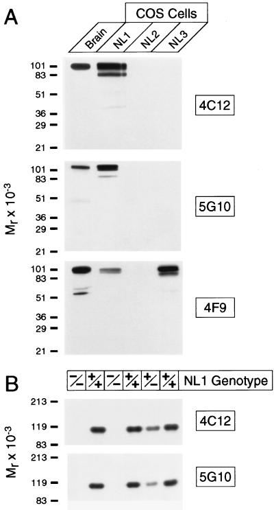
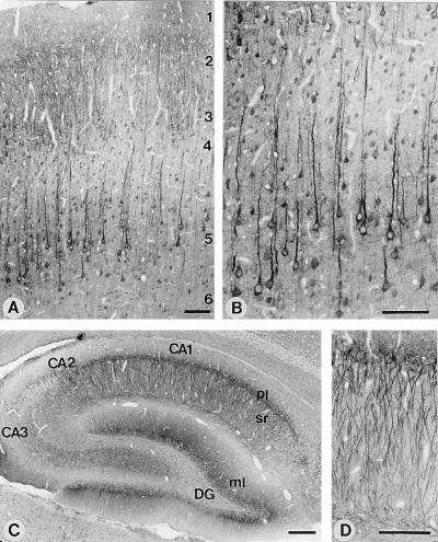
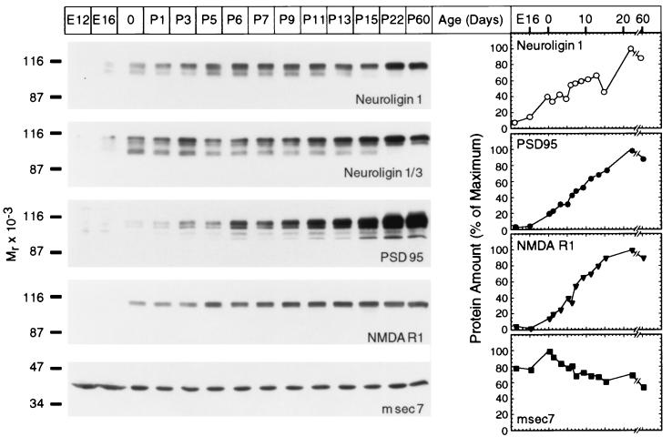
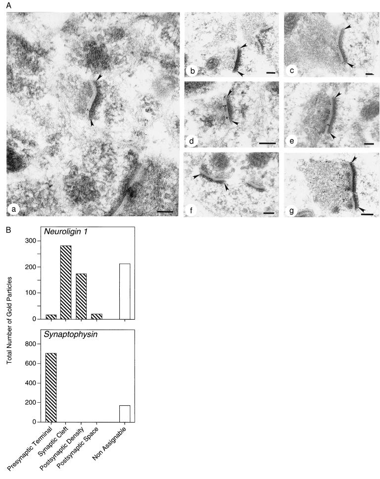
At the synapse, presynaptic membranes specialized for vesicular traffic are linked to postsynaptic membranes specialized for signal transduction. The mechanisms that connect pre- and postsynaptic membranes into synaptic junctions are unknown. Neuroligins and beta-neurexins are neuronal cell-surface proteins that bind to each other and form asymmetric intercellular junctions. To test whether the neuroligin/beta-neurexin junction is related to synapses, we generated and characterized monoclonal antibodies to neuroligin 1. With these antibodies, we show that neuroligin 1 is synaptic. The neuronal localization, subcellular distribution, and developmental expression of neuroligin 1 are similar to those of the postsynaptic marker proteins PSD-95 and NMDA-R1 receptor. Quantitative immunogold electron microscopy demonstrated that neuroligin 1 is clustered in synaptic clefts and postsynaptic densities. Double immunofluorescence labeling revealed that neuroligin 1 colocalizes with glutamatergic but not gamma-aminobutyric acid (GABA)ergic synapses. Thus neuroligin 1 is a synaptic cell-adhesion molecule that is enriched in postsynaptic densities where it may recruit receptors, channels, and signal-transduction molecules to synaptic sites of cell adhesion. In addition, the neuroligin/beta-neurexin junction may be involved in the specification of excitatory synapses.


Figures

Figure 1
Figure 1
Specificity of mAbs to neuroligin. (A) Immunoblots of rat brain homogenates and COS cells expressing neuroligins 1, 2, and 3 (10 μg per lane) probed with mAbs 4C12, 5G10, and 4F9. (B) Immunoblots of mouse brain homogenates from homozygous (−/−) and heterozygous (+/−) neuroligin 1-mutant mice and wild-type control mice (+/+).
Figure 2
Figure 2
Light-microscopic localization of neuroligin 1 in rat brain. Shown are sections of the overall neocortex (A) or of layers 4 and 5 of the neocortex (B). Numbers indicate cortical layers. Low-magnification view of the entire hippocampal formation (C) or high-magnification view of the apical dendrites of pyramidal neurons from the CA1 region (D). DG, dentate gyrus; ml, molecular layer; pl, pyramidal cell layer; and sr, stratum radiatum. All sections were stained with antibody 4C12 and developed by using horseradish peroxidase-labeled secondary antibodies. [Bar = 100 μm (A, B, and D) or 250 μm (C).]
Figure 3
Figure 3
Expression of neuroligin 1 in development. Brain homogenates from rats of the indicated age were analyzed by immunoblotting with antibodies to neuroligins, PSD-95, NMDA-receptor type 1 (NMDAR1), and msec7. Expression levels were quantified with iodinated secondary antibodies at the following ages: E12 and E16, embryonic days 12 and 16; 0, day of birth; P1–P60, postnatal days 1–60.
Figure 4
Figure 4
Quantitative immunogold electron microscopy of neuroligin 1 and synaptophysin 1. (A) Representative ultrathin sections from rat neocortex stained with antibodies to neuroligin 1 (af) and synaptophysin (g). Neuroligin 1 immunoreactivity decorates synaptic clefts (ae) and postsynaptic densities (f). Arrowheads indicate postsynaptic densities. (Bars = 0.1 μm.) (B) Quantitation of gold particles in randomly chosen sections labeled with antibodies to neuroligin 1 or synaptophysin. Total number of gold particles identified in the indicated subcellular compartments are given. Gold particles at locations that are not recognizable were designated nonassignable.
Figure 5
Figure 5
Confocal micrographs of brain sections double labeled for neuroligin 1 and GluR2/3 glutamate receptors (AI) or for GABAAβ receptors and glutamate receptors (JR). Note the colocalization of GluR2/3 receptors (a marker for excitatory synapses) with neuroligin 1 in somata and dendrites of pyramidal neurons in neocortex and hippocampus and in Purkinje cells of the cerebellum (AI). GABAAβ receptors, in contrast, are segregated (JR). (Bar = 100 μm.)

References

    1. Goodman, C. S. & Shatz, C. J. (1993) Cell72, Suppl., 77–98. - PubMed
    1. Goodman C S. Annu Rev Neurosci. 1996;19:341–377. - PubMed
    1. Chiba A, Keshishian H. Dev Biol. 1996;180:424–432. - PubMed
    1. Uchida N, Honjo Y, Johnson K R, Wheelock M J, Takeichi M. J Cell Biol. 1997;135:767–779. - PMC - PubMed
    1. Inoue A, Sanes J R. Science. 1997;276:1428–1431. - PubMed

Publication types

MeSH terms

LinkOut - more resources