Neuroretinal rim width ratios in morphological glaucoma diagnosis
- PMID: 9930265
- PMCID: PMC1722465
- DOI: 10.1136/bjo.82.12.1366
Neuroretinal rim width ratios in morphological glaucoma diagnosis
Abstract
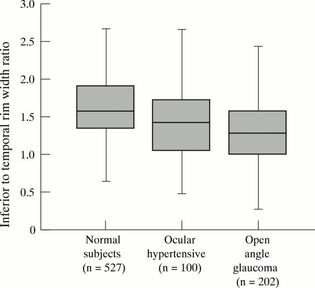
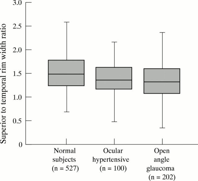
Aims: To evaluate the inferior to temporal neuroretinal rim width ratio and superior to temporal rim width ratio as measures of rim shape for diagnosis of glaucoma.
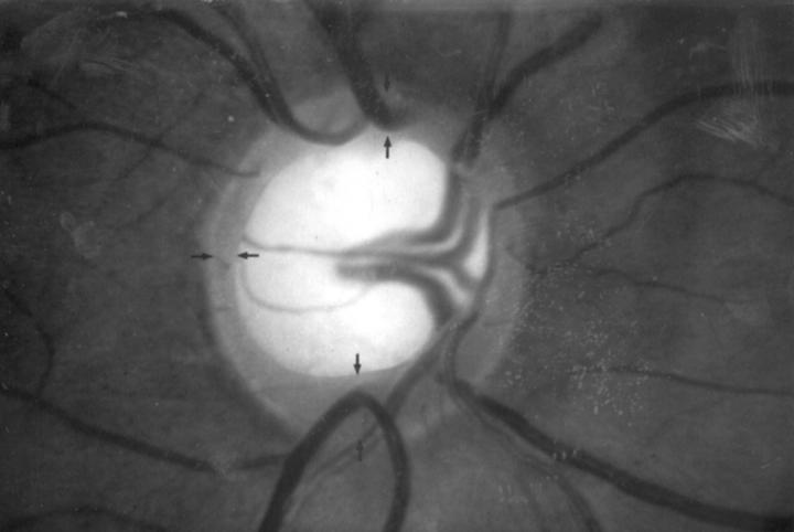
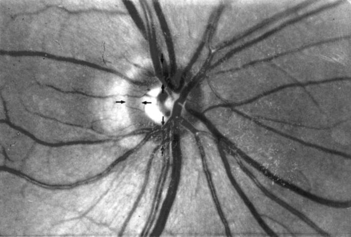
Methods: Colour stereo optic disc photographs of 527 normal subjects, 100 ocular hypertensive individuals with normal visual fields, and 202 open angle glaucoma patients with a mean perimetric defect of less than 10 dB were morphometrically evaluated. Eyes with an optic cup area of < 0.2 mm2 were excluded.
Results: In the normal subjects, inferior to temporal rim width ratio (1.67 (SD 0.53)) was significantly (p < 0.0001) higher than superior to temporal rim width ratio (1.56 (0.49)). Both ratios were significantly (p < 0.0001) higher the more vertically the optic disc was configured. In the normal eyes, both ratios were statistically independent of disc size, rim area, refractive error, age, and sex. With the differences being more marked for the inferior to temporal ratio than for the superior to temporal ratio, both rim width ratios were significantly (p < 0.005) lower in the ocular hypertensive group than in the normal group. Despite the high significance of the differences, diagnostic power of the inferior ratio and the superior ratio was 59% and 58%, respectively, indicating a marked overlap between the groups.
Conclusions: Abnormally low inferior to temporal and superior to temporal rim width ratios can indicate glaucomatous optic nerve damage in some ocular hypertensive eyes. Being independent of optic disc size and ocular magnification, the rim width ratios may be taken as one among other variables for the ophthalmoscopic optic disc evaluation, taking into account, however, a pronounced overlap between normal eyes and ocular hypertensive eyes.
Figures


Similar articles
-
Ranking of optic disc variables for detection of glaucomatous optic nerve damage.Invest Ophthalmol Vis Sci. 2000 Jun;41(7):1764-73. Invest Ophthalmol Vis Sci. 2000. PMID: 10845597
-
Comparison of measurements of neuroretinal rim area between confocal laser scanning tomography and planimetry of photographs.Br J Ophthalmol. 1998 Apr;82(4):362-6. doi: 10.1136/bjo.82.4.362. Br J Ophthalmol. 1998. PMID: 9640181 Free PMC article.
-
The optic disc hemifield test.J Glaucoma. 2004 Apr;13(2):108-13. doi: 10.1097/00061198-200404000-00005. J Glaucoma. 2004. PMID: 15097255
-
The morphology of the optic nerve head in the Singaporean Chinese population (the Tanjong Pagar study): part 1--Optic nerve head morphology.Br J Ophthalmol. 2008 Mar;92(3):303-9. doi: 10.1136/bjo.2007.123273. Br J Ophthalmol. 2008. PMID: 18303151 Review.
-
Spotlight on the Disc-Damage Likelihood Scale (DDLS).Clin Ophthalmol. 2021 Oct 7;15:4059-4071. doi: 10.2147/OPTH.S284618. eCollection 2021. Clin Ophthalmol. 2021. PMID: 34675474 Free PMC article. Review.
Cited by
-
Glaucoma screening in Western Cameroon.Graefes Arch Clin Exp Ophthalmol. 2009 Dec;247(12):1671-5. doi: 10.1007/s00417-009-1166-7. Epub 2009 Aug 23. Graefes Arch Clin Exp Ophthalmol. 2009. PMID: 19701764
-
Optic disk size and glaucoma.Surv Ophthalmol. 2007 Jan-Feb;52(1):32-49. doi: 10.1016/j.survophthal.2006.10.002. Surv Ophthalmol. 2007. PMID: 17212989 Free PMC article. Review.
-
Pattern Electroretinogram Parameters Are Associated with Optic Nerve Morphology in Preperimetric Glaucoma after Adjusting for Disc Area.J Ophthalmol. 2021 Oct 13;2021:8025337. doi: 10.1155/2021/8025337. eCollection 2021. J Ophthalmol. 2021. PMID: 34845426 Free PMC article.
-
Optic disc morphology in south India: the Vellore Eye Study.Br J Ophthalmol. 2003 Feb;87(2):189-96. doi: 10.1136/bjo.87.2.189. Br J Ophthalmol. 2003. PMID: 12543749 Free PMC article. Clinical Trial.
-
Anatomy and evaluation of the optic nerve head.Arq Bras Oftalmol. 2022 Feb 14;85(6):636-643. doi: 10.5935/0004-2749.20220080. eCollection 2022. Arq Bras Oftalmol. 2022. PMID: 35170637 Free PMC article. Review.
References
Publication types
MeSH terms
LinkOut - more resources
Full Text Sources
Medical
Miscellaneous